
What is an MRI simulator?
Software that reproduces an MRI scanner's console without the magnet, a magnet-free mock scanner that prepares children, or a radiotherapy planning scanner. The types, how they work, the evidence, and what to look for.
By the ScanFlo Editorial Team · Updated
In this section · Simulation
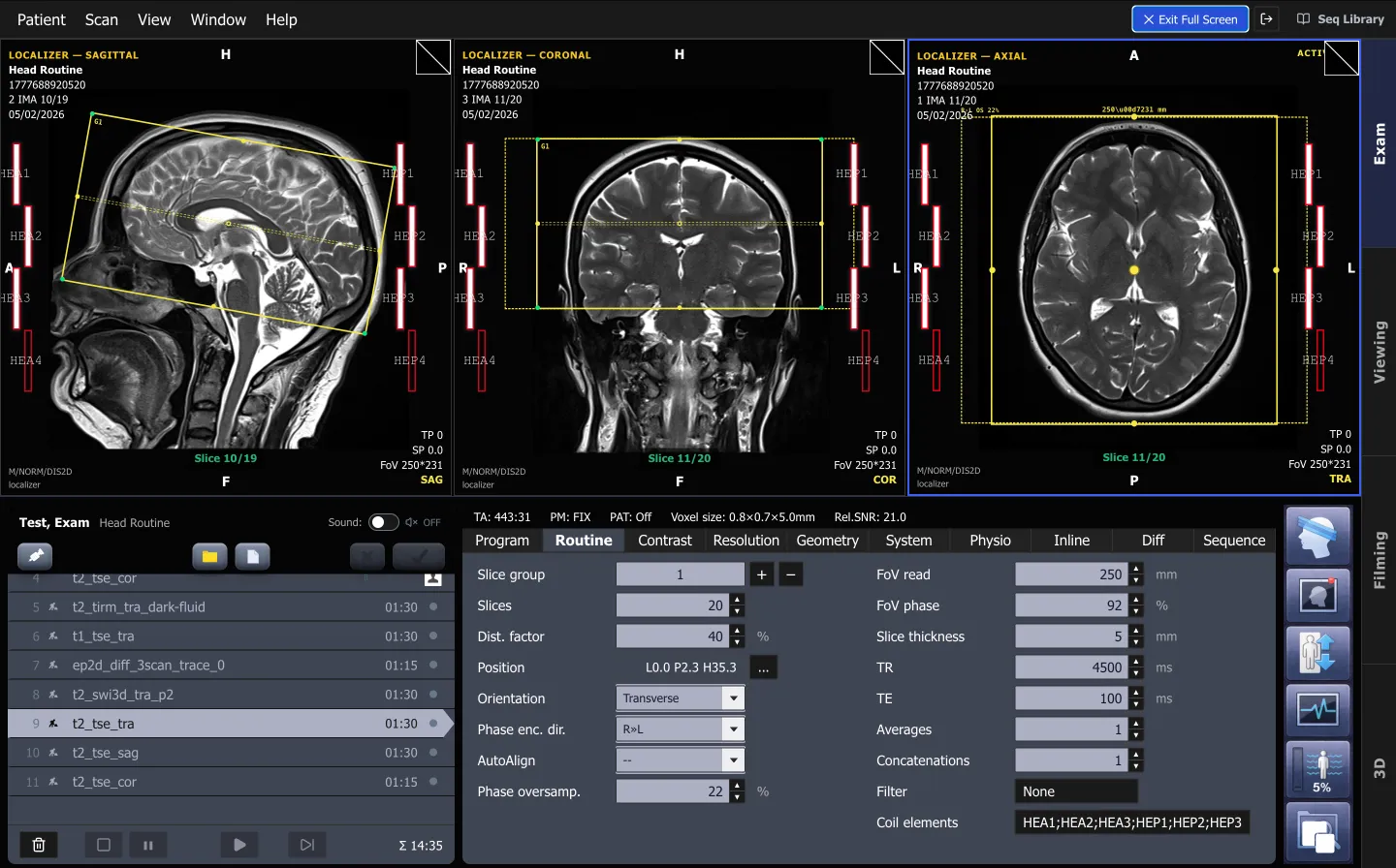
An MRI simulator is software that reproduces an MRI scanner's console without the magnet. You plan slices on localizers, choose sequences, set TR, TE, flip angle and resolution, and get an image that responds the way a real acquisition would. The same name is also used for magnet-free mock scanners that prepare children for MRI, and for scanners used to plan radiotherapy.
Sources: Carter et al. (2010); Sensors (2021); Chaka and Hardy (2021); McDaid et al. (2021).
Three things called an MRI simulator
The phrase covers three different tools, and search results mix them up. Know which one you need.
What people mean by 'MRI simulator'
The radiotherapy meaning is the easiest to rule out: Cahill and colleagues describe implementing "a retrofit MRI Simulator for RT planning", which is a working scanner, not a teaching tool (Cahill et al., 2023).
Does a mock MRI scanner reduce sedation in children?
Yes. Several studies found that a practice session in a mock scanner lets most young children have their real scan awake.
Children scanned without anaesthesia after mock scanner preparation
The effect is largest in the youngest children. Carter and colleagues found the anaesthesia rate was 16.8% lower in children aged 3 to 8 who had a mock MRI (Carter et al., Pediatric Radiology, 2010). It does not need expensive kit, either: a multi-centre study found no difference between a commercial simulator and "an inexpensive mock scanner consisting of a toy tunnel, vibrating mat, and video player" (Barnea-Goraly et al., 2014).
What types of MRI training simulator exist?
Four software families, which differ in how they make the image: image banks, signal-equation simulators, full Bloch-equation engines and VR experiences.
| Type | How the image is made | Strength | Limitation |
|---|---|---|---|
| Image bank / console replica | Shows stored images for the chosen protocol | Realistic console workflow | Images do not change when parameters change |
| Signal-equation (formula) | Computes each tissue's signal from TR, TE and its T1, T2 and PD | Instant, runs in a browser, good for contrast teaching | Simplified physics; artefacts must be modelled separately |
| Bloch-equation engine | Simulates spins voxel by voxel, fills k-space and reconstructs | Physically accurate, used for sequence research | Compute-heavy; usually code-driven, not a console |
| VR experience | A 3D room for positioning, safety or patient preparation | Spatial skills and rehearsal | Narrow scope; needs headsets |
Bloch engines are mostly open-source research tools. JEMRIS, the Jülich Extensible MRI Simulator, was published in 2010 (Stöcker et al.); MRiLab reported a speed-up of about 200 times by running on a GPU (Liu et al., 2017); KomaMRI, from 2023, solves the Bloch equations with CPU and GPU parallelisation and matched JEMRIS to within 0.1% (Castillo-Passi et al., 2023). They are excellent for physicists and poor for teaching console workflow, because they are driven by code rather than a scanner interface.
How a teaching simulator turns your settings into an image
- STEP 01Patient modelTissue maps with T1, T2 and proton density for every voxel.
- STEP 02Your protocolSequence, TR, TE, flip angle, slice, field of view, matrix.
- STEP 03SignalEach tissue's signal computed from the protocol and its properties.
- STEP 04ArtefactsWrap, motion or chemical shift added when the plan invites them.
- STEP 05ImageContrast, noise and scan time update as you change settings.
Does training on an MRI simulator improve learning?
The evidence is positive and still young. Randomised and curriculum studies show gains in knowledge and exam scores, while reviews ask for more rigorous validation.
In a randomised pre- and post-test study of 60 learners at a hospital in Madrid, the group that used an MRI simulator scored significantly higher on both the post-test and the gain (Treceño-Fernández et al., Sensors, 2021). The authors note why simulators matter: busy departments give "minimum (or null) scanner time allocation for training". A 2026 programme that redesigned 40 hours of a 204-hour MRI course around a simulator saw final exam means rise from 17.55 to 18.34 out of 20 (Vafapour et al., Academic Radiology, 2026).
Simulation should complement, rather than replace, supervised MRI exposure, particularly for patient positioning, coil handling, in-room safety checks, and other tactile or psychomotor competencies.
A review of 663 articles on computer-based simulation in CT and MRI education found simulators used mainly for scan planning, acquisition and reconstruction, and concluded that their educational validation "has not been fully assessed through rigorous academic assessments and metrics" (Chaka and Hardy, Radiography, 2021). Our guide on whether simulator practice transfers to the scanner goes through that evidence in detail.
What should you look for in MRI training software?
Parameters that really change the image, planning on real localizers, artefacts on demand, and access that scales to a whole batch.
An educator's checklist
That list follows the design requirements set out in the Sensors 2021 study, which named protocols, planning, editable timing parameters, artefacts at the trainer's demand, k-space, trainer roles, speed and easy access among them (Treceño-Fernández et al., 2021).
ScanFlo MRI · exam console
Can a simulator teach MRI safety?
Partly. Software and e-learning teach the rules and the screening logic well; in-room checks still need supervised practice.
An MR safety e-learning module, built for a radiotherapy department, raised scores from 59% to 79% (McDaid et al., JMIRS, 2021), and the authors note that guidance suggests annual safety training for everyone working in MRI. In a survey of radiography students, 69% said they would use skills learned in simulation in clinical practice (Elshami and Abuzaid, JMIRS, 2017).
Where a simulator fits in a curriculum
Try it on the console
Try an MRI simulator in your browser
ScanFlo MRI runs on real localizers, with TR, TE, TI and flip angle driving the image live. Free for students.
Start free →Related: the radiography simulation guide, TR and TE, and MRI protocol coverage.
Frequently asked questions
Is an MRI simulator the same as an MRI phantom?
No. A phantom is a physical test object scanned on a real MRI scanner, usually for quality control. An MRI simulator is software that produces images without a scanner, or, in paediatrics, a magnet-free mock scanner used to prepare patients.
Do I need a powerful computer to use an MRI simulator online?
Not for a teaching simulator. Browser-based teaching simulators run on an ordinary laptop; the SimMRI authors note theirs computes entirely on the client, which suits large student groups. Research engines that solve the Bloch equations for every spin are the ones that benefit from a GPU.
Can a home-made mock scanner work as well as a commercial one?
In one multi-centre study of children, success rates did not differ between centres using a commercial MRI simulator and those using an inexpensive mock scanner made from a toy tunnel, a vibrating mat and a video player.
Which children benefit most from a mock MRI?
Carter and colleagues found the largest effect in children aged 3 to 8, where the general anaesthesia rate was 16.8% lower than in children who did not have a mock scan.
Can an MRI simulator show artefacts?
A good teaching simulator should. Generating acquisition artefacts on the trainer's demand, such as wrap, motion or chemical shift, was one of the design requirements in the Sensors 2021 study, because diagnosing and fixing them is a core technologist skill.
Does simulator practice count as clinical hours?
That is decided by your university or registration body, not by the software. Treat simulator time as preparation for clinical postings, and check your programme's rules before logging it.
Sources
Every figure on this page links back to one of these.
- Carter AJ, et al. Mock MRI: reducing the need for anaesthesia in children. Pediatric Radiology, 2010
- Mork-Knudsen H, et al. Pediatric brain MRI without sedation: optimization by simulation. BMC Pediatrics, 2026
- de Bie HMA, et al. Preparing children with a mock scanner training protocol results in high quality structural and functional MRI scans. European Journal of Pediatrics, 2010
- Barnea-Goraly N, et al. High success rates of sedation-free brain MRI scanning in young children using simple subject preparation protocols with and without a commercial mock scanner. Pediatric Radiology, 2014
- Treceño-Fernández D, et al. Magnetic resonance simulation in education: quantitative evaluation of an actual classroom experience. Sensors, 2021
- Chaka B, Hardy M. Computer based simulation in CT and MRI radiography education: current role and future opportunities. Radiography, 2021
- Vafapour H, et al. Curriculum-integrated MRI simulation across successive cohorts. Academic Radiology, 2026
- McDaid L, et al. Developing electronic learning to deliver MR safety training in a radiotherapy department. JMIRS, 2021
- Elshami W, Abuzaid M. Transforming magnetic resonance imaging education through simulation-based training. JMIRS, 2017
- Cahill K, et al. Implementation of a retrofit MRI simulator for radiation therapy planning. Journal of Medical Radiation Sciences, 2023
- Stöcker T, et al. High-performance computing MRI simulations (JEMRIS). Magnetic Resonance in Medicine, 2010
- Liu F, et al. Fast realistic MRI simulations based on generalized multi-pool exchange tissue model (MRiLab). IEEE Transactions on Medical Imaging, 2017
- Castillo-Passi C, et al. KomaMRI.jl: an open-source framework for general MRI simulations with GPU acceleration. Magnetic Resonance in Medicine, 2023
- SimMRI, a browser-based MRI simulator for teaching. ISMRM 2023, abstract 2396