
PET-CT acquisition types: step and shoot, continuous bed motion and static
A PET ring sees 15 to 26 cm of the patient; a whole body needs about a metre. Step and shoot, continuous bed motion and static acquisition are three ways round that, and knowing how many beds a patient needs, and why, is where planning starts.
By the ScanFlo Editorial Team · Updated
In this section · PET-CT fundamentals
The ring is short because detectors are expensive, and every extra centimetre of axial coverage costs more crystal and electronics. How PET-CT works covers what the ring detects. What follows is how the table carries the patient through it: in steps, continuously, or not at all.
Step and shoot
The table parks, the ring counts for a set time, the table advances and the ring counts again. Each parking place is a bed position. This is the routine whole-body method on most scanners and the one most exam cards are written around.
The table never advances by a full field of view. In 3D PET the ring is most sensitive at its axial centre and least sensitive at the two edges, because a photon pair born near an edge has fewer detector pairs that can catch it. If beds were placed end to end, the images would carry a band of higher noise at every join. So consecutive beds overlap and share their weakest slices. Overlap is usually somewhere between a quarter and a half of the field of view, depending on the scanner and the reconstruction. ScanFlo defaults to 23.5%.
Overlap is also why the bed count is not simply length divided by ring length. Each bed adds only the part of the field of view it does not share with its neighbour:
beds = (scan length − overlap) ÷ (field of view − overlap), rounded up
Take a skull-base to mid-thigh range of about 1,000 mm on a 218 mm ring at 23.5% overlap. The overlap is 51 mm, each bed advances 167 mm, and (1,000 − 51) ÷ 167 comes to 5.7, which becomes six beds. The same patient on a 261 mm ring needs five. A tall patient, or a range extended to include the legs, adds beds; a paediatric patient removes them.
Two practical consequences follow. The planning box on the topogram snaps: you cannot ask for exactly 1,000 mm, only for a whole number of beds, and the range becomes whatever those beds cover. And scan time is bed count multiplied by time per bed. Time per bed is set by the protocol, typically a couple of minutes on older systems and less on digital ones. For heavy patients, the usual guidance is to lengthen the time per bed rather than push the injected activity higher. Many sites also give the beds over the region of interest longer than the rest.
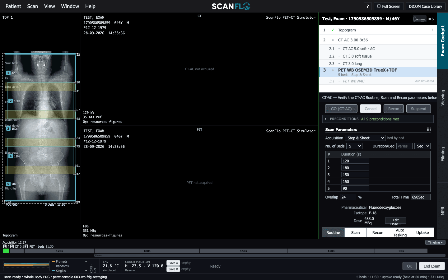
Direction matters too. Some departments start at the thighs and scan upwards, so that the pelvis is imaged soon after the patient has voided and before the bladder refills. Others start at the head. Either way, the patient lies with arms raised for body imaging where they can manage it. Arms by the side add attenuation and truncation artefacts across the chest and abdomen.
Continuous bed motion
Continuous bed motion takes away the stops. The table glides through the ring at a controlled speed while the scanner acquires the whole time, so every point in the range passes through the full axial sensitivity profile. The ScanFlo console calls it Continuous bed motion.
Removing the stops removes the snapping. The range starts and ends exactly where it is drawn, to the millimetre, and there are no bed boundaries to plan around. The more useful change is that speed becomes a parameter. The range can be split into segments, each with its own speed: slow over the liver or pelvis where the clinical question is, faster over the legs where counts matter less. Duration for each segment is simply its length divided by its speed, so a 400 mm segment at 1 mm/s takes 400 seconds. ScanFlo allows up to four ranges between 0.4 and 10 mm/s.
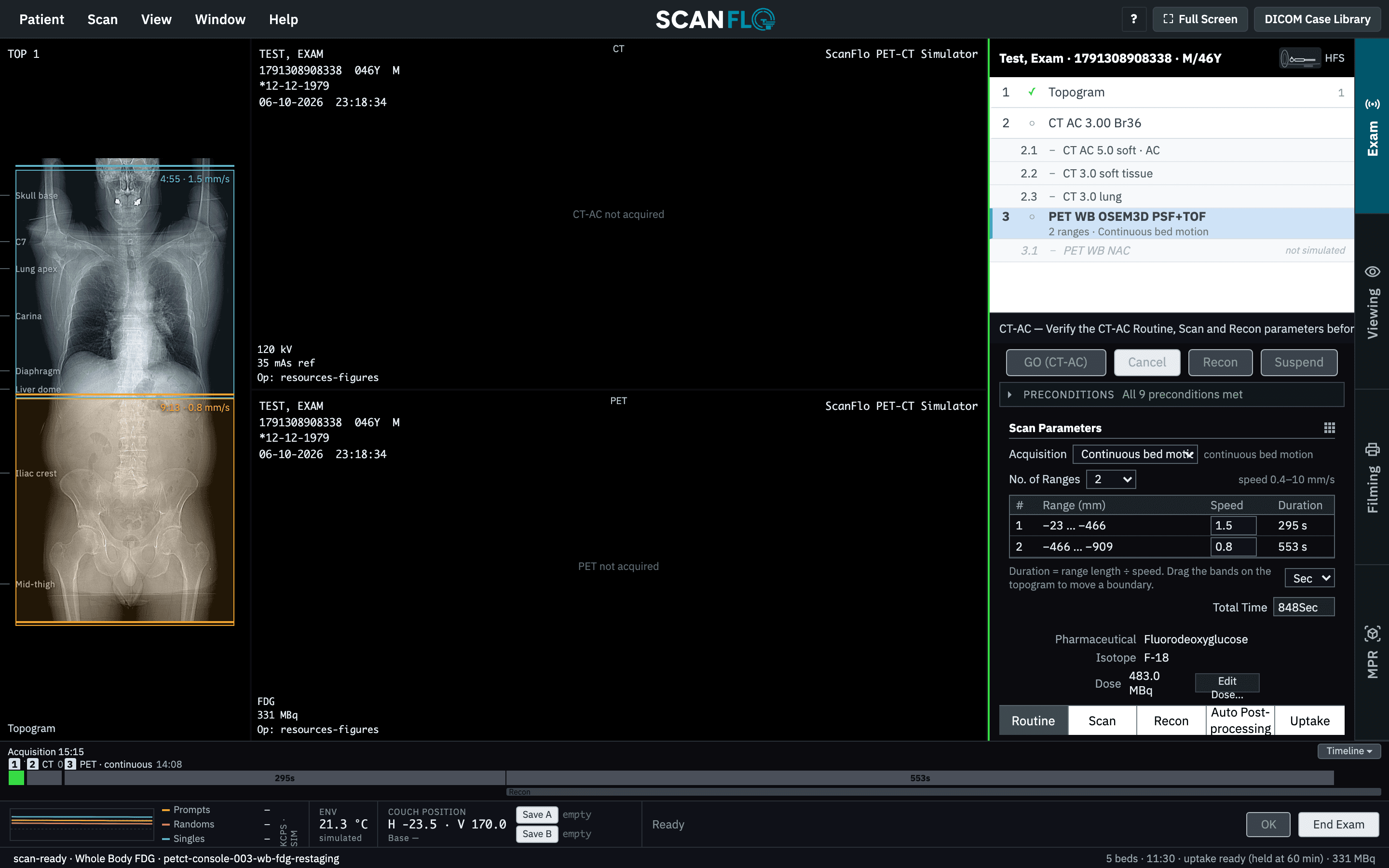
What continuous motion does not do is create counts from nothing. For the same total time and the same injected activity, the statistics are broadly comparable to step and shoot. The gain is in how that time is spent: noise is more uniform along the patient, coverage matches the anatomy, and time goes where the question is.
Static
Sometimes the whole question fits inside one field of view. Brain FDG, myocardial perfusion and a single joint or region are all acquired with the bed parked at one position for the whole acquisition. There is no overlap to set and no range to snap. The only planning decision is where to centre the ring, and on the heart that decision deserves care, because a heart pushed to one edge of the field of view sits in the ring's weakest slices.
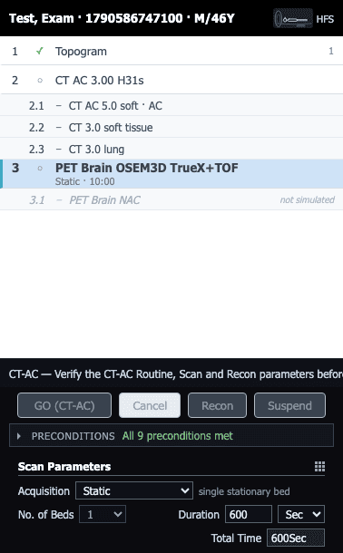
Static acquisitions are also where timing changes shape. A whole-body scan waits for the tracer to settle and then takes a snapshot. A static acquisition can start at the moment of injection and record in list mode, so the data can later be split into a series of frames showing the tracer arriving and washing out. This is how myocardial blood flow is measured with N-13 ammonia, whose ten-minute half-life leaves no time to wait anyway.
Choosing between them
| Mode | Table | Coverage | Typical use |
|---|---|---|---|
| Step & Shoot | Stops at overlapping beds | Whole beds; the range snaps | Routine whole-body FDG, PSMA, DOTATATE |
| Continuous bed motion | Moves continuously | Exact range, up to 4 speed segments | Whole-body with a slow segment over the region of interest |
| Static | Parked at one position | One field of view | Brain, cardiac perfusion, dynamic studies |
The tracer usually decides first. FDG, PSMA and DOTATATE are whole-body questions, so they get step and shoot or continuous motion. N-13 ammonia is a heart question, so it gets a static bed. NaF looks for bone disease that may be anywhere in the skeleton, so its range often runs further down the legs than an oncology study. PET radiotracers explains why.
Try it in ScanFlo: Plan a whole-body FDG study in Step & Shoot and drag the lower edge of the emission box down the thighs. Watch it jump in whole-bed steps while No. of Beds and Total Time follow. Then change Acquisition to Continuous bed motion, set the number of ranges to two and give the range over the trunk a slower speed than the one over the legs. Compare the total time with the bed plan you started from. See ScanFlo PET-CT →