
Diffusion-weighted imaging
DWI measures how far water can move in a few tens of milliseconds. Where it cannot move, as in an acute infarct or a packed tumour, the image stays bright. Read it with the ADC map every time, or T2 shine-through will fool you.
By the ScanFlo Editorial Team · Updated
In this section · MRI fundamentals
What DWI measures
Water molecules wander. In a glass of water they wander freely; in tissue they bump into membranes, fibres and crowded cells. How far they get depends on how the tissue is built, which is why a single diffusion sequence can pick up a stroke within minutes of onset and grade the cellularity of a tumour.
The sequence adds two strong, matched gradient pulses either side of the refocusing pulse. A stationary proton is dephased by the first and rephased by the second and keeps its signal. A proton that has moved in between sees a different field the second time, is not fully rephased and loses signal. So in a diffusion-weighted image, tissue where water moves freely goes dark and tissue where water is trapped stays bright. The physics is set out in MRI physics: from protons to pictures.
The b-value
How strongly the image is weighted is expressed as the b-value, in s/mm², which combines gradient strength, duration and spacing. At b=0 there is no diffusion weighting and the image is essentially T2-weighted. As b rises, freely moving water fades and only restricted water keeps its signal. The cost is signal-to-noise ratio, which falls quickly at high b.
| Region | Common b-values (s/mm²) | Why |
|---|---|---|
| Brain | 0 and 1000 | Stroke, abscess, cellular tumour |
| Body (liver, lymph nodes) | 50 and 800 | Low b for lesion detection, higher for restriction |
| Prostate | Up to 1400–2000 | Suppress background gland so tumour stands out |
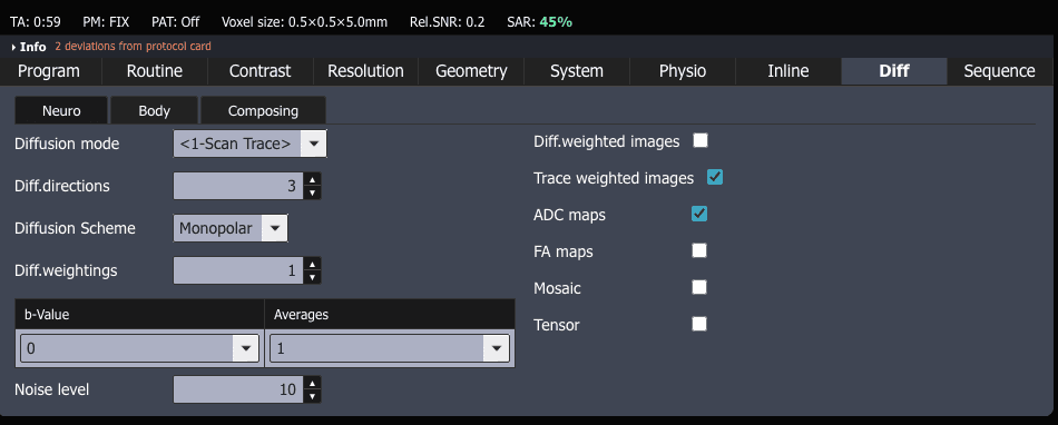
Very high b-values are often computed from lower acquired ones instead of scanned directly, which saves time and noise. ScanFlo can do this: the "Computed b-value images" option on the diffusion card.
Always read it with the ADC map
A bright diffusion image is not proof of restriction, because the image still carries its T2 weighting. A lesion with very long T2 can glow on b=1000 while its water moves perfectly freely. This is T2 shine-through, and it is the classic trap.
The apparent diffusion coefficient map removes it. ADC is calculated from the signal at two or more b-values and represents diffusion alone. Free water has a high ADC and is bright on the map; restricted water has a low ADC and is dark. True restriction is bright on DWI and dark on ADC. Shine-through is bright on both. Read the two together every time.
Where it earns its place
In the brain, acute ischaemia restricts within minutes, long before CT or T2 shows anything, because failed cell pumps let water flood into swelling cells. DWI also separates an abscess, whose thick pus restricts, from a necrotic tumour, whose liquid centre usually does not, and it identifies epidermoid cysts. In the body, restriction tracks cellularity. It helps detect liver metastases and involved nodes, leads prostate MRI in the peripheral zone and is used to follow treatment response.
Why the images look the way they do
Nearly all clinical DWI uses echo-planar imaging, which reads a whole slice after a single excitation and freezes the patient's motion against the large diffusion gradients. EPI is fast and fragile. It distorts and loses signal near air and bone: the frontal sinuses, the temporal lobes and the rectum. Parallel imaging shortens the readout and reduces this. Where distortion matters most, readout-segmented or reduced field of view techniques trade time for fidelity.
Try it in ScanFlo: Run the diffusion sequence on a brain case and open the b=1000 trace image next to the ADC map. CSF should be dark on the trace and bright on ADC, which is what free water looks like. If the case carries a lesion, use the pair to decide whether it truly restricts. Then change the b-value and watch the background fade. See ScanFlo MRI →