
PET radiotracers: FDG, NaF, NH3, PSMA and DOTATATE
The scanner counts the same photons whatever is injected. The tracer decides the question, the patient preparation, the uptake time and what normal looks like.
By the ScanFlo Editorial Team · Updated
In this section · PET-CT fundamentals
The same patient can have an FDG scan on Monday and a PSMA scan on Thursday and look like two different people. Many reading errors in PET come from judging one tracer by another tracer's normal, so the sections below say what should be bright before anything is called abnormal.
| Tracer | Isotope (half-life) | What it follows | Typical uptake before scan |
|---|---|---|---|
| FDG | F-18 (110 min) | Glucose metabolism | About 60 min |
| NaF | F-18 (110 min) | Bone remodelling | About 30–60 min |
| N-13 ammonia | N-13 (10 min) | Myocardial blood flow | None; acquire from injection |
| PSMA | Ga-68 (68 min) or F-18 | PSMA on prostate cancer cells | About 60 min for Ga-68; often longer for F-18 agents |
| DOTATATE | Ga-68 (68 min) | Somatostatin receptors | About 60 min |
Uptake times vary between guidelines and departments. What matters most for comparison is keeping the uptake time consistent between a baseline and a follow-up scan.
FDG: glucose, and everything that competes with it
Fluorodeoxyglucose is taken into cells as if it were glucose and phosphorylated, and then it stops. It cannot continue down the metabolic pathway, so it stays trapped in proportion to how hungry the cell is for glucose. Many cancers are very hungry, and so are inflammation and infection, which is why FDG does both oncology and a fair amount of infection imaging.
Nearly all of the preparation exists to stop other tissues from competing. The patient fasts, typically for four to six hours, because a meal raises insulin and insulin pushes FDG into muscle and fat. Blood glucose is checked before injection against the department's threshold. The day before, strenuous exercise is avoided. During uptake the patient rests quietly and warm, without talking or reading much, because working muscles take up tracer and cold switches on brown fat in the neck and shoulders. The scan follows about an hour after injection.
Normal FDG is busy. The brain is intensely avid. The myocardium ranges from invisible to blazing depending on what the patient last ate. The kidneys and bladder carry excreted tracer, the bowel is patchy, and the liver provides the background that other organs are compared against. A hot spot needs to be read against that pattern before it is called disease.
NaF: where bone is remodelling
Sodium fluoride is the simplest tracer here. Fluoride ions are exchanged into the bone mineral wherever blood flow and remodelling are high. A metastasis provoking new bone takes it up, but so does a healing fracture or an arthritic joint. NaF is sensitive and not specific, and it is read with the CT beside it for exactly that reason.
No fasting is needed; good hydration helps clear the soft-tissue background. Imaging usually starts somewhere between 30 and 60 minutes after injection. Because the disease can be anywhere in the skeleton, the range often extends further down the legs than an oncology study, sometimes to the feet.
N-13 ammonia: the heart, on a ten-minute clock
Ammonia is extracted by heart muscle in proportion to blood flow, which makes it a perfusion tracer. Its half-life of about ten minutes rules the logistics. It has to come from a cyclotron close to the scanner, the patient is positioned before the dose arrives, and the acquisition begins at injection. It is a static acquisition over the heart, recorded in list mode so that dynamic frames can be extracted for flow quantification. A full study compares rest with pharmacological stress. The short half-life is what allows the second injection to follow soon after the first.
ScanFlo currently includes one N-13 ammonia study, acquired at rest, and uses it to teach polar-map reading. There is no stress study yet.
PSMA: a target on prostate cancer
Prostate-specific membrane antigen is a protein that prostate cancer cells express in large amounts. PSMA ligands bind it, which gives staging and recurrence imaging a sensitivity that neither CT nor bone scan could reach. PSMA imaging is also how patients are selected for lutetium-177 PSMA therapy, because the same target carries the therapeutic dose.
The isotope changes the practicalities. Ga-68 PSMA-11 is made from a generator, has a 68-minute half-life and is usually scanned at about an hour. F-18 agents last about 110 minutes, can be shipped from a cyclotron, and are often scanned later, up to around two hours. Excretion differs too. PSMA-11 goes out through the urine, so the bladder is bright next to the prostate bed. PSMA-1007, the F-18 agent ScanFlo uses, is cleared mainly through the liver, which quietens the bladder and raises the liver background. Normal uptake in the salivary and lacrimal glands, kidneys and small bowel is expected with all of them. No fasting is required.
DOTATATE: somatostatin receptors
Well-differentiated neuroendocrine tumours carry somatostatin receptors, subtype 2 in particular, and Ga-68 DOTATATE is a peptide built to bind them. It is the workhorse for neuroendocrine tumours and, like PSMA, the gatekeeper for radionuclide therapy with lutetium-177 DOTATATE.
The preparation is about the patient's own drugs. Octreotide and lanreotide bind the same receptors and can block the tracer, so the scan is usually booked just before the next long-acting injection is due, and short-acting octreotide is stopped for about a day. Imaging follows at about an hour. Normal DOTATATE lights up the spleen most of all, then the kidneys, liver, adrenals and pituitary. The uncinate process of the pancreas often shows focal uptake that is physiological and easily mistaken for a tumour.
From tracer to acquisition
Once the tracer is known, most of the acquisition follows. FDG, PSMA and DOTATATE are whole-body questions, answered with step and shoot or continuous bed motion from the skull base to mid-thigh after about an hour. NaF uses the same whole-body approach over a longer range. N-13 ammonia is a static bed over the heart that starts with the injection. PET-CT acquisition types covers how each of those is planned.
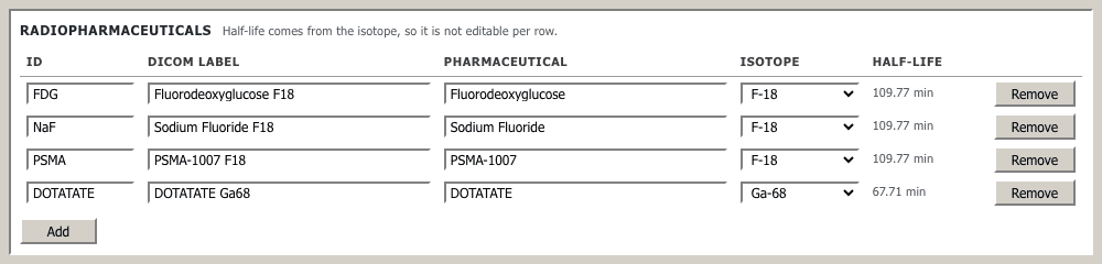
Try it in ScanFlo: Open Whole Body FDG from the protocol list, then Whole Body PSMA, Bone NaF and Whole Body DOTATATE in turn. Read the pharmaceutical and isotope on each routine card, and open Edit Dose to see the half-life the decay correction will use. If your case library holds studies for more than one tracer, open their MIPs side by side and, before looking for disease, name every bright organ and why it should be bright. See ScanFlo PET-CT →