
Resources/Imaging informatics
DICOM explained for radiographers
Every image you produce leaves the console as a DICOM object. Understanding what that object contains — and how it travels — is the difference between operating a scanner and running an imaging department.
What DICOM is
DICOM — Digital Imaging and Communications in Medicine — is the international standard that governs how medical images are stored, transmitted, retrieved, printed and displayed. It was developed jointly by the American College of Radiology and the National Electrical Manufacturers Association, and it is the reason a scanner from one manufacturer can send images to an archive from another and a workstation from a third can open them.
Two things are worth separating early, because people conflate them. DICOM is a file format: a container that binds the image to its clinical and technical context. It is also a network protocol: a defined conversation devices have when they exchange those files. Radiographers meet both every shift — the first when reviewing a study, the second every time a series fails to reach PACS.
Why standardisation mattered
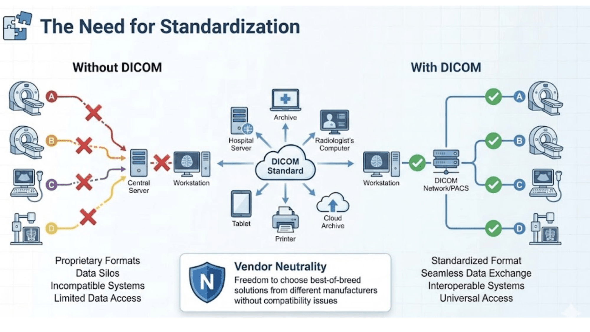
Before the standard settled, manufacturers wrote their own formats. Images were trapped on the console that produced them, departments bought whole ecosystems from a single vendor to keep anything working together, and moving a study between hospitals meant physical media and hope. Standardisation broke that: a department can now buy the best scanner, the best archive and the best reading workstation independently — vendor neutrality that only exists because the exchange format is agreed.
Anatomy of a DICOM object
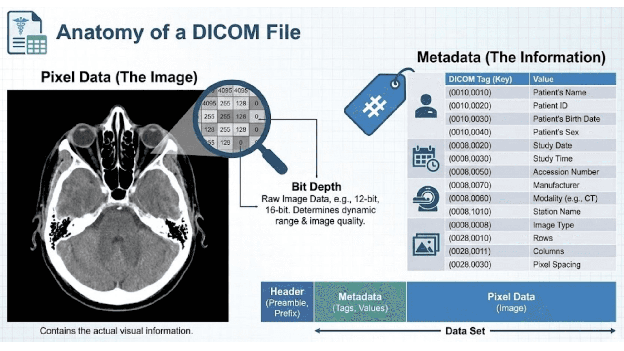
A DICOM file is not an image with a label attached. It is a data set in which the pixels are one element among many, each element identified by a numeric tag.
| Tag | Element | Why it matters |
|---|---|---|
| (0010,0010) | Patient's name | Identity — and the first thing anonymisation removes |
| (0010,0020) | Patient ID | The stable key linking studies over time |
| (0008,0020) | Study date | Chronology for comparison studies |
| (0008,0050) | Accession number | Links the image to the order in the hospital system |
| (0008,0060) | Modality | CT, MR, US, CR — drives how viewers display it |
| (0008,0070) | Manufacturer | Device provenance for troubleshooting |
| (0028,0010) | Rows | Image matrix height |
| (0028,0011) | Columns | Image matrix width |
| (0028,0030) | Pixel spacing | Physical size per pixel — without it, measurements are meaningless |
Alongside the metadata sits the pixel data itself, stored at a bit depth — commonly 12 or 16 bits — that determines how many distinct grey levels the image can represent. That dynamic range is why a CT dataset can be windowed for lung, soft tissue and bone from a single acquisition: the information is all present, and windowing chooses which slice of it to display.
The information hierarchy
DICOM organises data in four levels, and the model maps directly onto how imaging work is actually structured:
- Patient — the person, identified consistently across every visit.
- Study — one examination, typically one visit for one clinical question.
- Series — one acquisition within that study: a sequence, a reconstruction, a plane.
- Instance — one image within a series.
A routine brain MRI is one study containing perhaps eight to twelve series, each holding twenty to two hundred instances. Understanding this hierarchy is what makes efficient querying possible — and it is why sending "the scan" to a colleague is ambiguous until you say whether you mean the study, one series, or a single image.
Modalities the standard covers
DICOM spans CT, MRI, radiography, ultrasound, PET-CT, nuclear medicine and radiation therapy planning, among others. Each carries modality-specific attributes: a CT object records tube voltage, exposure and dose descriptors, while an MR object records repetition time, echo time, flip angle and sequence type. Same container, different contents — which is why a viewer can display any modality but only a modality-aware tool can interpret the acquisition parameters properly.
Networking: how images actually move
Devices on a DICOM network identify themselves with an Application Entity title, an IP address and a port. When something fails to arrive, those three values are the first place to look — a mismatched AE title is a far more common cause of a missing study than any fault in the image itself.
| Service | What it does | Everyday example |
|---|---|---|
| C-STORE | Sends an object to another device | Scanner pushing a completed series to PACS |
| C-FIND | Queries for studies matching criteria | Searching the archive by patient or date |
| C-MOVE / C-GET | Retrieves objects | Pulling a prior study to a workstation |
| C-ECHO | Verifies connectivity | The DICOM equivalent of a ping when a link is suspect |
Where it meets the department
- PACS — the picture archiving and communication system: centralised storage, retrieval and distribution of every study the department produces.
- Modality worklist — scheduled examinations pushed from the information system to the scanner, so the technologist selects a patient instead of typing one. This single mechanism removes most demographic errors at their source.
- Storage commitment — an explicit confirmation from the archive that it has taken permanent responsibility for the images, so a console can safely clear local storage.
The practical consequence for a radiographer is worth stating plainly: selecting the wrong worklist entry mislabels a study, and a mislabelled study is harder to correct after it reaches the archive than any technical error you might make during the scan itself.
Where the standard is heading
- DICOMweb — web-based access to studies over standard HTTP, which is what makes browser-based viewers and cloud archives practical.
- AI and machine-learning integration — algorithms consuming DICOM objects and writing results back as structured objects rather than screenshots.
- Cloud storage — archives that scale without a server room, with the same standard governing access.
- Security — TLS-encrypted transport, access control, and de-identification workflows that strip patient identifiers when images are used for teaching or research.
That last point deserves emphasis for anyone building teaching material: de-identification is not optional. Removing the visible name from a screenshot does nothing about the dozens of identifying tags inside the object.
Common questions
- What does DICOM stand for?
- Digital Imaging and Communications in Medicine. It is the international standard that defines both the file format medical images are stored in and the network protocol devices use to exchange them.
- Is DICOM a file format or a network protocol?
- Both, and that dual nature is the point. The format keeps the image and its patient, study and acquisition metadata in one object, while the protocol defines how scanners, PACS, workstations and printers talk to each other.
- What is the difference between a study, a series and an instance?
- A study is one imaging examination for a patient. A series is one acquisition within it — a single sequence or reconstruction. An instance is one image within that series. A brain MRI study might hold ten series, each with dozens of instances.
- Why can't a DICOM image just be saved as a JPEG?
- A JPEG carries pixels and nothing else. A DICOM object carries the pixel data plus who was scanned, when, on which device, with which parameters, at what slice position and spacing — the context that makes the image diagnostic and traceable.
- What is a modality worklist?
- A list of scheduled examinations pushed from the hospital information system to the scanner, so the technologist selects the patient rather than typing details in. It removes the single most common source of mislabelled studies.
- What does C-STORE mean?
- It is the DICOM service that sends an image object from one device to another — typically scanner to PACS. Related services include C-FIND for querying, C-MOVE and C-GET for retrieval, and C-ECHO for connectivity testing.
Try it in ScanFlo: After your next simulated exam, look at what the study contains before filming it: how many series did the protocol produce, and what distinguishes each one? Naming the series correctly in your head is the same skill that keeps a real archive searchable.
Related: The console · MRI protocol coverage