
How to get hands-on MRI practice before your first clinical posting
ScanFlo Editorial Team · · 8 min read
In this article
- What does an MRI student actually do on a first posting?
- How long is the MRI posting in a B.Sc. MRIT internship?
- What are the four MRI safety zones?
- Why can't a student walk into the scan room?
- How does MRI screening work, and how can you practise it?
- Can an MRI simulator replace scanner time?
- A four-week practice plan before your posting
You get hands-on MRI practice before your first posting by rehearsing everything that does not need a patient: the safety zones, the screening form, cross-sectional anatomy and, on an MRI simulator, slice planning and parameters. The posting itself is short. Arriving with those habits is what turns two months of watching into two months of doing.
Most students meet the magnet for the first time on their internship, with a radiographer, a patient and a waiting list all present at once. It is a lot to absorb. This guide covers what the posting looks like in India, what the safety rules allow a student to do, and a four-week plan for the time before it starts.
What does an MRI student actually do on a first posting?
Mostly observe, help with screening and positioning, and log what you see. Running a scan on your own comes later, under supervision.
That isn't only an Indian experience. A study of radiography students at the University of Malta found MRI placements were mainly based on observation
, with students lacking hands-on practice. The same students said helping the team with patient screening is what built their confidence (Portanier Mifsud, Castillo and Portelli, Radiography, 2015).
Indian syllabi set similar expectations. JIPMER's B.Sc. curriculum expects interns to be able to assist senior technicians in performing MRI examinations
, and lists planning head and body studies and MRI safety instruction among the practicals (JIPMER, 2021).
How long is the MRI posting in a B.Sc. MRIT internship?
Under the NCAHP model curriculum it is 2 months of a 1-year, 2,400-hour rotating internship. Your university's own rules decide the final number.
Colleges don't all follow one pattern, so check your own handbook before you plan around a number.
| Programme | Internship | MRI posting |
|---|---|---|
| NCAHP model curriculum | 1 year, 2,400 hours | 2 months |
| RK University | 1 year, at least 1,440 hours | 2 months |
| JIPMER | 1 year, compulsory | Rotation within the year |
| SGRR University | 6 months, compulsory | Rotation within the six months |
What are the four MRI safety zones?
The American College of Radiology divides an MRI suite into four zones, from the public corridor to the magnet room itself, with access tightening at each step.
Indian departments write their own local rules, but most are built on this layout, and it is the vocabulary your supervisors will use.
Paraphrased from the ACR Manual on MR Safety (2024). The magnet is always on, so a pair of scissors in a pocket is a projectile in Zone IV whether a patient is on the table or not.
Why can't a student walk into the scan room?
Because until you finish your facility's MR safety training, the rules treat you as non-MR personnel, and non-MR personnel go into Zone IV only under supervision.
The ACR defines non-MR personnel as anyone who has not completed the facility's formal MR safety education within the previous 12 months
. It also recommends annual refresher training for everyone working in the suite (ACR, 2024). Ask about that training in your first week. It is the gate to everything else.
How does MRI screening work, and how can you practise it?
Every patient fills a written screening form, is screened twice (at least once by trained MR staff), and gets a final check from the technologist before entering Zone IV.
You can rehearse this tonight. Get a copy of your hospital's screening form, or any published one, and run through it with a classmate playing the patient. Swap roles. Then do it again with a patient who has a pacemaker card in their wallet and forgets to mention it.
Prepare for nerves too. In a survey of 50 patients at a hospital in Ambala, Haryana, 30% reported fear before their MRI and only 20% reported no worry at all (Sharma and Singh, IJRMS, 2023). A calm two-sentence explanation of noise, time and the call bell does more than any technique.
Can an MRI simulator replace scanner time?
No. It complements it, letting you rehearse the console decisions so that your supervised scanner time goes on judgement and patient care.
The evidence is promising and still small. In a randomised study of 60 students in Madrid, the group taught with an MRI simulator scored significantly higher after training than the group taught without it (Treceño-Fernández and colleagues, Sensors, 2021). One study is not a guarantee, and nobody has yet measured how simulator-trained students perform on a real scanner months later.
A four-week practice plan before your posting
One theme a week: safety, anatomy, protocols, then putting it together. Ninety minutes a day is enough.
On day one of the posting
Try it on the console
Plan your first scan before your posting
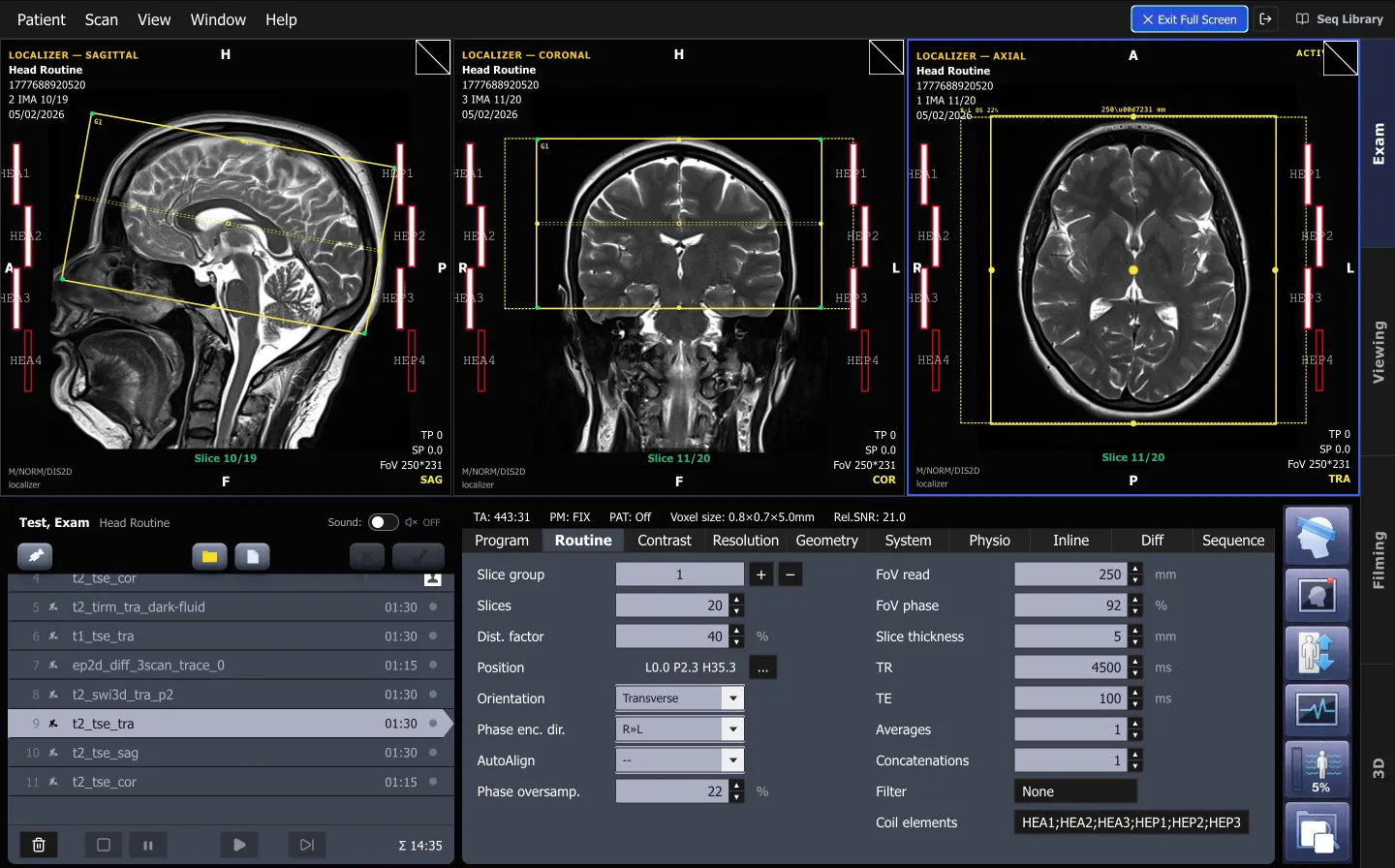
ScanFlo MRI runs in a browser on real localizers. Plan a brain, change TR and TE, and see the image respond.
Start free →Related reading: what an MRI simulator is, how colleges teach MRI without scanner time, and brain MRI protocols.
Sources
Every figure in this article links back to one of these.
- American College of Radiology. ACR Manual on MR Safety, 2024
- NCAHP model B.Sc. Medical Radiology and Imaging Technology curriculum, as adopted by VNSGU, 2026
- RK University. Guidelines for internship training, B.Sc. MRIT
- JIPMER. B.Sc. Medical Radiology Imaging Technology curriculum, 2021
- SGRR University. BMRIT board of studies, 2023-24
- Portanier Mifsud C, Castillo J, Portelli JL. Radiography students' clinical placement experiences in MRI: a phenomenological study. Radiography, 2015
- Treceño-Fernández D, et al. Magnetic resonance simulation in education: quantitative evaluation of an actual classroom experience. Sensors, 2021;21(18):6011
- Sharma SK, Singh S. Knowledge and perception of magnetic resonance imaging safety among patients. Int J Res Med Sci, 2023;11(11):4088-94
Frequently asked questions
Do I need deep MRI physics before my first posting?
No. The NCAHP model curriculum puts MRI technique in the sixth semester, and many students reach their posting with only the basics. What helps more is knowing the safety zones, the screening form, the standard brain, spine and knee protocols, and cross-sectional anatomy in three planes.
Is the B.Sc. MRIT internship paid?
The NCAHP model curriculum sets a minimum stipend of Rs 7,000 a month during the internship. What colleges and hospitals actually pay varies, so ask your department.
What should I remove before entering the MRI scan room?
Anything metallic or magnetic: phone, keys, coins, hairpins, belts, watches, bank and ID cards, pens and scissors. You will also be screened in writing before you enter. Follow your department's checklist, which is the authority on site.
What goes in an MRI log book?
Each examination you saw or assisted: date, body part, protocol, your role, and any event worth recording. RK University's internship guidelines, for example, ask interns to record every event of each posting in a log book.
Can I practise MRI scanning at home without a scanner?
Yes, with a browser-based MRI simulator. ScanFlo MRI lets you plan slices on localizers, change TR, TE and flip angle, and see the image respond, on a laptop. It is practice for the console decisions, not a substitute for supervised scanning.