
How PET-CT works
PET shows where a molecule went. The CT shows where that is, and tells the PET how much signal was lost on the way out. Between them they produce a number, the SUV, that is only as good as the details recorded at the front desk.
By the ScanFlo Editorial Team · Updated
In this section · PET-CT fundamentals
What the scanner counts
A PET tracer is an ordinary molecule with a positron-emitting atom attached. Once injected, the molecule goes wherever its chemistry sends it: glucose to cells that burn glucose, fluoride to bone that is remodelling, a peptide to the receptor it was built to fit. The scanner never sees the molecule. It sees what happens when the attached atom decays.
The emitted positron travels a millimetre or so, meets an electron and the two annihilate. Their mass becomes two photons of 511 keV that leave in almost exactly opposite directions. A ring of detectors around the patient waits for pairs: two hits on opposite sides within a few nanoseconds are counted as one event, and the annihilation must have happened somewhere on the line joining those two detectors. Collect enough of these lines and reconstruction turns them into a three-dimensional map of activity.
That map is quantitative, which is what separates PET from most of the imaging a radiographer does. The grey level of a CT pixel describes attenuation. The value of a PET voxel is meant to be a concentration in becquerels per millilitre, and everything downstream, from the SUV to the response assessment, assumes that number is right.
Why the CT is not optional
The number is only right after correction. A photon pair born in the liver has to cross far more tissue than one born in a superficial lymph node, and many of them never reach the ring. Left alone, the reconstruction would show the centre of the body as colder than it is and the skin as a bright rim.
The CT supplies the fix. Its attenuation values are converted from the energy of the X-ray beam to the 511 keV of annihilation photons, and the resulting map tells the reconstruction how much signal each line of response lost on its way out. This is why the CT in a PET-CT is often acquired at modest dose. For attenuation correction and localisation it does not need to be diagnostic, although many departments run a full diagnostic CT when the referral asks for one.
Because the correction trusts the CT completely, anything wrong with the CT is copied into the PET. Breathing is the classic case. The CT freezes the diaphragm in one position in a second or two, while the PET averages it over minutes, so a lesion at the lung base can land on the wrong side of the dome and be corrected with the wrong attenuation. Dense metal and concentrated oral or IV contrast push the correction the other way and can manufacture uptake. When a hot spot sits at an interface or next to metal, look at the non-attenuation-corrected images before believing it. Real uptake is still there without the correction; a correction artefact disappears.
SUV, and who controls it
Concentration alone would be hard to compare between patients, since a heavier patient given the same dose dilutes it more. The standardised uptake value normalises for that: tissue concentration divided by injected activity per kilogram. Tissue with an SUV of 1 holds exactly its share of the dose. A tumour with an SUV of 12 holds twelve times its share.
Look at what goes into that fraction. Body weight, taken at the desk. Injected activity, which is the syringe reading minus whatever stayed in the syringe and line. Injection time, because the tracer has been decaying since then. Uptake time, because uptake keeps rising for longer than most protocols wait. Blood glucose, for FDG. Every one of these is recorded by a radiographer or a technologist, not by the scanner, and a mistake in any of them changes the SUV without changing the image at all. An extravasated injection is the worst of them: part of the dose sits in the arm and the whole-body values fall.
Two normalisations appear on most workstations. SUVbw uses total body weight. SUVlbm, sometimes written SUL, uses lean body mass, which behaves better in heavy patients because fat takes up little FDG and inflates body-weight SUV. Whichever your department reports, use the same one at baseline and follow-up.
A ring shorter than the patient
One physical fact shapes the rest of the examination. The detector ring has an axial field of view of roughly 15 to 26 cm on most clinical systems. The scanner modelled in ScanFlo covers 218 mm; the longest clinical rings reach about 26 cm. A whole-body PET runs from the skull base to mid-thigh, about a metre, so the ring has to be moved along the patient. How it moves is the subject of PET-CT acquisition types. How long you wait before moving it at all depends on the tracer, which is covered in PET radiotracers.
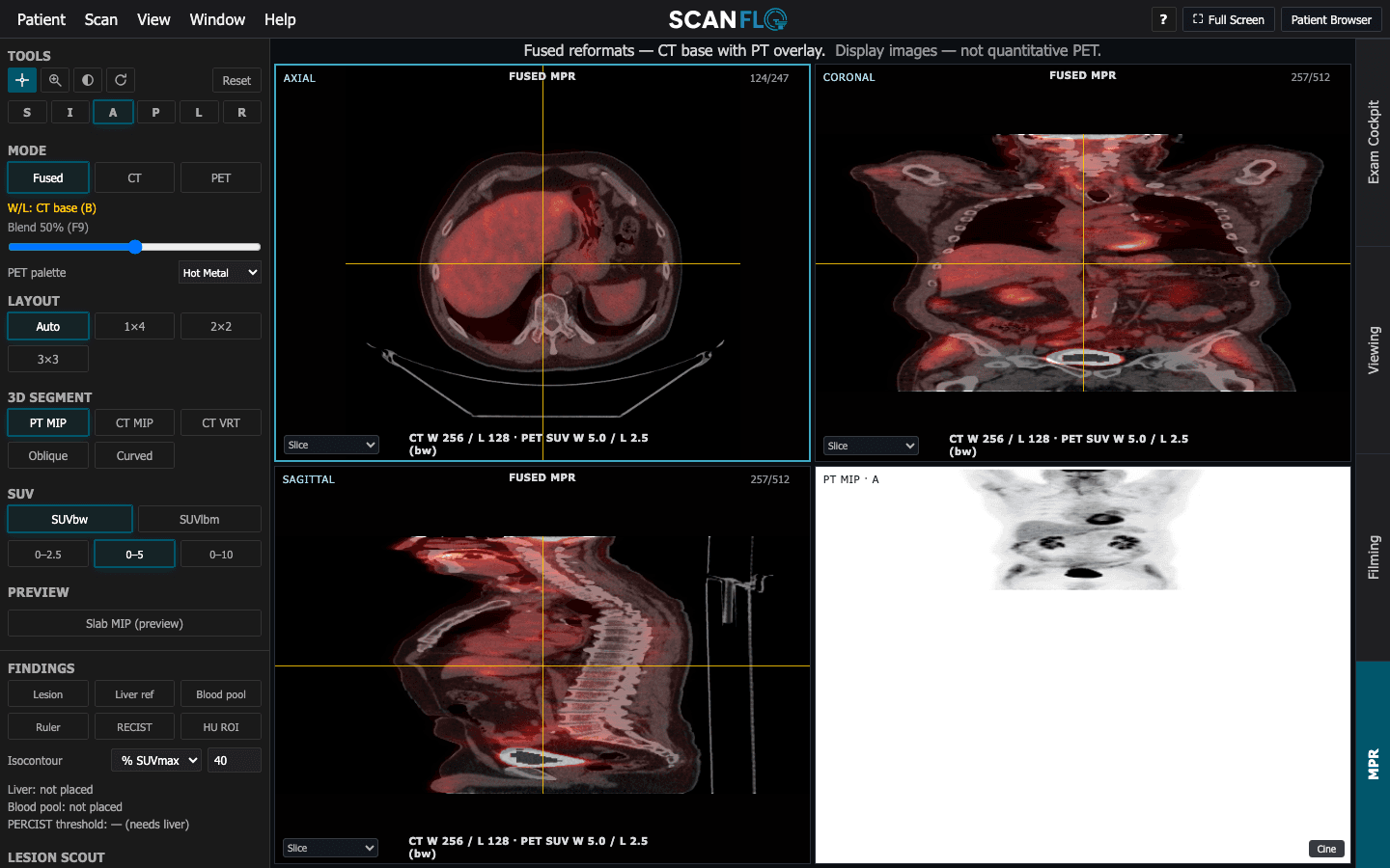
Try it in ScanFlo: Open an FDG case, register the patient and run the exam through to the fused images. In MPR, place the liver reference and switch between SUVbw and SUVlbm. The liver is the usual reference organ; if its SUV looks implausible, check the weight and injection time on the registration card before blaming the scanner. See ScanFlo PET-CT →