
Teaching MRI without scanner time: how colleges use simulators
ScanFlo Editorial Team · · 8 min read · Updated
In this article
- Why is scanner time so hard to get for MRI students?
- What is simulation-based radiography education?
- Does the evidence show students learn from simulators?
- How much simulation do other countries recommend?
- What does India's NCAHP curriculum say about skills labs?
- Where does an MRI simulator fit in the semester?
- How should faculty run a simulation session?
Colleges teach MRI without scanner time by moving everything that does not need a patient onto a simulator: slice planning, parameter choices, artefacts and safety screening. Students then use their scarce posting hours for the parts only a real patient can teach. This is what simulation-based radiography education means in practice.
Every radiography educator in India knows the arithmetic. One scanner, a long patient list, and a batch of students who need to learn it. The scanner earns its keep scanning people. Practice sessions come last.
Why is scanner time so hard to get for MRI students?
Because scanners are scarce and fully booked. A government research centre puts India at one and a half to two MRI machines per million people.
Machines are only part of the gap. As Guy Frija, professor emeritus of radiology in Paris, told RSNA News, radiology in low- and middle-income countries needs specific spaces for equipment, stable power, regular maintenance and future upgrades
(RSNA, 2024). A teaching slot on that machine is the easiest thing to cut.
What is simulation-based radiography education?
Learning scanning tasks in a setting where mistakes cost nothing and any step can be repeated: software simulators, phantoms, role-play and VR.
For MRI, a software simulator does most of the work, because the skills are decisions at a console. Where should the slice group go? What happens to contrast if TR doubles? Which artefact is that, and what would you change? None of those needs a magnet.
Does the evidence show students learn from simulators?
Yes, for knowledge and short-term skills. Randomised studies found simulator-trained groups beat their controls, though none has measured what happens to patients.
| Study | What was compared | Finding |
|---|---|---|
| Sensors, 2021 | 60 students in Madrid, MRI simulator vs teaching without it | Simulator group scored significantly higher after training |
| Sapkaroski et al., 2019 | 76 first-year students, VR training vs role-play in a real clinical room, tested 3 weeks later | VR group 36% better on digit separation, 23% on central ray position |
| Partner et al., 2022 | A one-week simulation package in place of placement | Students' perceptions supported it as a replacement for one week of placement |
| Scoping review, 2024 | 78 radiography simulation studies | Skills most often measured (66 studies); no patient outcomes reported |
Read those numbers honestly. A review in Radiography found that evidence for replacing clinical time comes from small student samples
(Jimenez and colleagues, 2023). Simulation prepares students for postings. It does not replace them.
How much simulation do other countries recommend?
The UK's College of Radiographers recommends about 120 hours of simulation per student, on top of 1,200 hours of practice-based learning. In the US, ARRT lets ten imaging procedures be simulated.
The conversation around the use of simulation-based education (SBE) within diagnostic radiography education and training has shifted from should it be used to how we use it effectively.
What does India's NCAHP curriculum say about skills labs?
The competency-based MRIT curriculum lists skills laboratory as a teaching method and asks institutes to provide simulation and skill labs in the early years of studentship.
The exact line is Provide simulation and skill labs for practicing skills specific to the program in the initial years of observership/studentship
(NCAHP curriculum, Gurugram University edition). Careers360 reported that the new curricula would be rolled out mandatorily
from 2026 (Careers360, 2025). For a college, that turns a skills lab from a nice-to-have into something an inspector can ask about.
Where does an MRI simulator fit in the semester?
Before the first posting for planning, protocols and safety, then alongside postings for the sequences and pathology students rarely see.
How should faculty run a simulation session?
Use the same five-step cycle the UK simulation model recommends, and tie each session to one written learning outcome.
Measure the result. A student who enjoyed the session has not necessarily learned anything. Pre- and post-tests, or a short OSCE station, tell you whether the hour was worth it.
Try it on the console
Run a simulation lab without buying a scanner
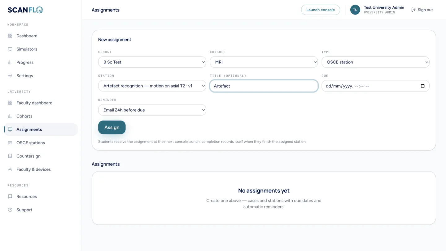
ScanFlo gives each student an MRI, CT and PET-CT console in the browser. Faculty set assignments and OSCE stations and see who completed them.
See ScanFlo for colleges →Related: the radiography simulation guide, ScanFlo for universities, and whether simulator practice transfers to the scanner.
Sources
Every figure in this article links back to one of these.
- Inter-University Accelerator Centre. Indigenous MRI project
- World Health Organization. Global Health Observatory, indicator DEVICES08: MRI units per million population
- RSNA News. Imaging in low- and middle-income countries, July 2024
- NCAHP competency-based curriculum, Bachelor of Medical Radiology and Imaging Technology (Gurugram University edition)
- Careers360. NCAHP competency-based curricula for allied healthcare, 2025
- College of Radiographers. Position statement on practice-based learning, July 2025
- Wilkinson E, et al. The role and efficacy of simulation in pre-registration education and training of diagnostic radiographers. University of Bradford for the College of Radiographers, January 2025
- Vestbøstad M, Karlgren K, Olsen N. Research on simulation-based training in radiography: a scoping review. Cochrane Colloquium abstracts, 2024
- Treceño-Fernández D, et al. Magnetic resonance simulation in education: quantitative evaluation of an actual classroom experience. Sensors, 2021
- Sapkaroski D, Baird M, Mundy M, Dimmock MR. Quantification of student radiographic patient positioning using an immersive virtual reality simulation. Simulation in Healthcare, 2019;14(4):258-63
- Jimenez YA, et al. Can simulation-based education or other education interventions replace clinical placement in medical radiation sciences? A narrative review. Radiography, 2023;29(2):421-7
- Partner A, Shiner N, Hyde E, Errett S. First year student radiographers' perceptions of a one-week simulation-based education package designed to increase clinical placement capacity. Radiography, 2022;28(3):577-85
- ARRT. Radiography didactic and clinical competency requirements
Frequently asked questions
Do students need MRI physics before they use a simulator?
A little helps, but the simulator is a good place to learn it. Changing TR or TE and watching contrast change makes the physics lecture concrete. Start with planning and safety, and bring in parameters once students can place a slice group.
Can one simulator serve a whole batch?
With browser-based software, yes: each student works on their own laptop or a lab computer, so a batch of 20 can practise at the same time. Physical phantoms and VR headsets are shared and need a rota.
Does simulator time count towards studentship or log book hours?
That is decided by each university. The NCAHP curriculum asks for simulation and skill labs and lists skills laboratory as a teaching method, but how those hours are logged is set locally. Check with your board of studies.
Can simulation teach MRI safety screening?
Yes, and it is one of the best uses. Screening is a structured conversation that students can role-play with sample forms before they ever stand at the Zone III door.
Can the same approach work for CT and X-ray?
Yes. CT simulators teach topogram planning, dose and contrast timing, and X-ray simulators teach positioning and exposure without radiation. ScanFlo covers MRI, CT, PET-CT and X-ray in one browser platform.