
Abdomen X-ray positioning
How to position for each of the 10 abdomen X-ray views: where the patient goes, where to centre, which receptor and distance to use, and what a good image looks like.
By the ScanFlo Editorial Team · Updated
In this section · X-ray positioning
- X-ray positioning guides
- Skull positioning
- Face & sinuses positioning
- Soft tissue neck positioning
- C-spine positioning
- Chest positioning
- Shoulder positioning
- Humerus positioning
- Elbow positioning
- Forearm positioning
- Wrist positioning
- Hand positioning
- T-spine positioning
- Abdomen positioning
- L-spine positioning
- Sacrum & SI joints positioning
- Pelvis positioning
- Hip positioning
- Femur positioning
- Knee positioning
- Tibia & fibula positioning
- Ankle positioning
- Foot positioning
X-ray positioning/Abdomen
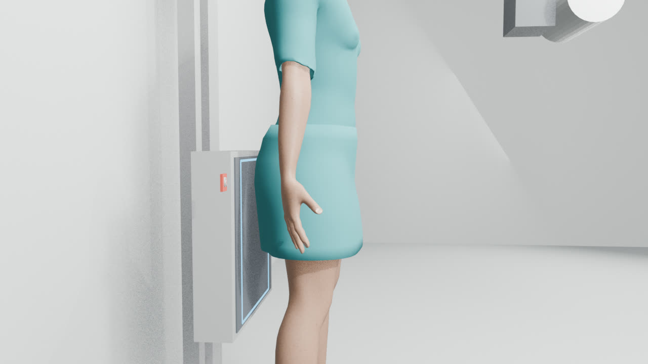
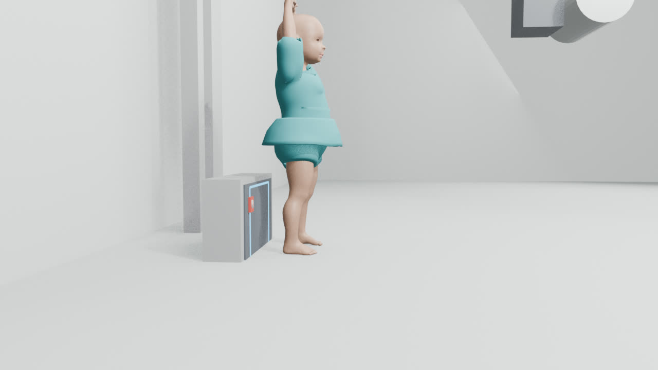
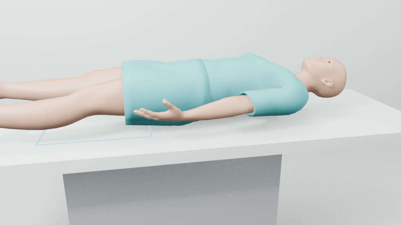
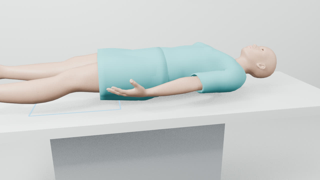
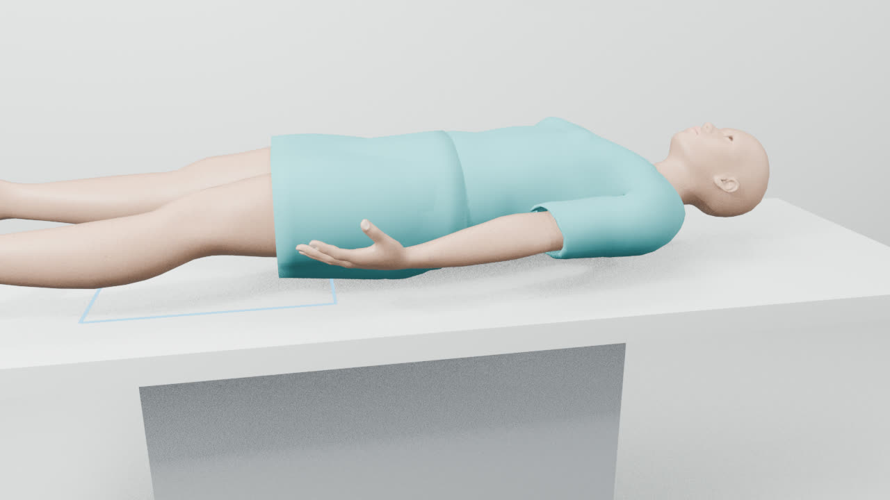
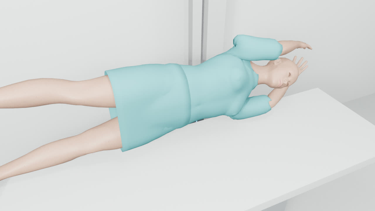
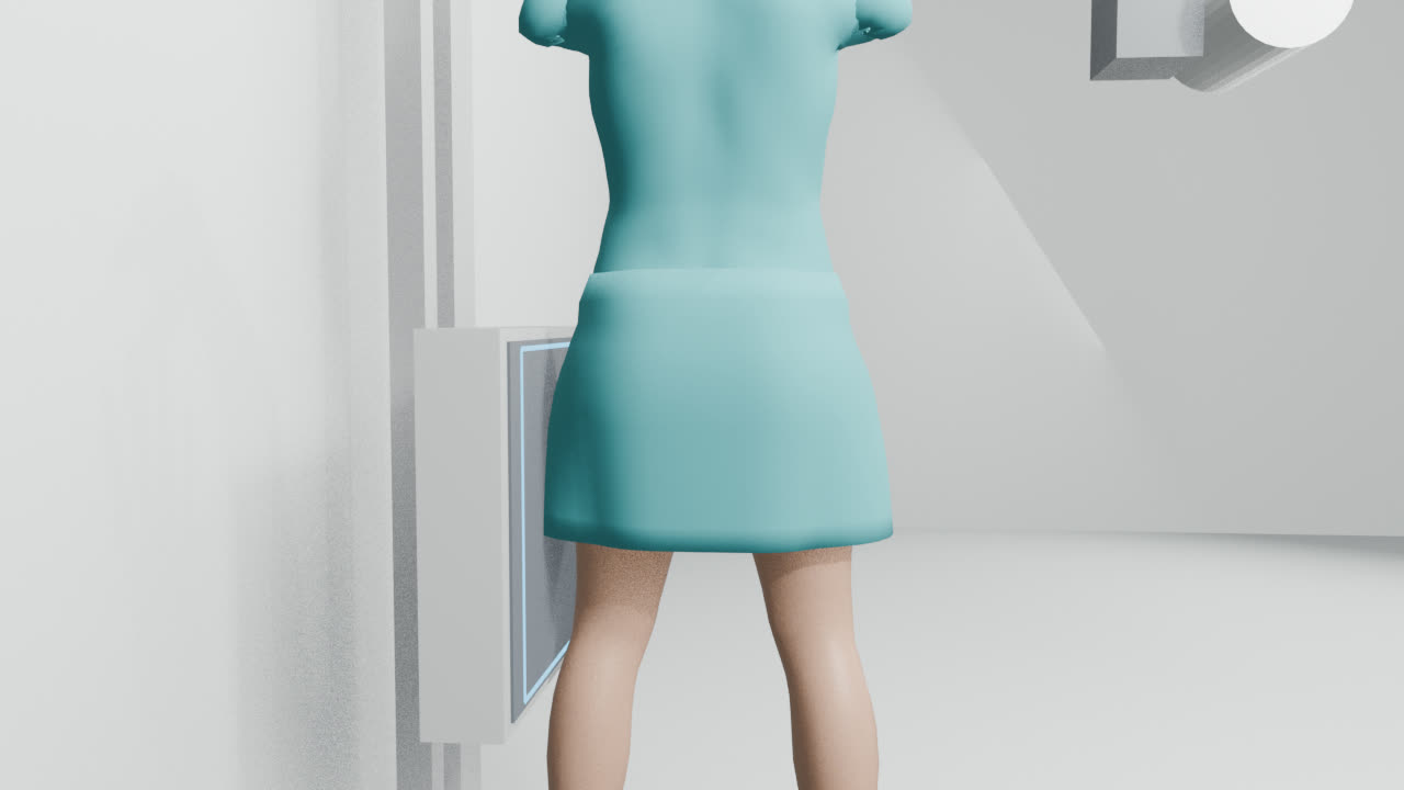
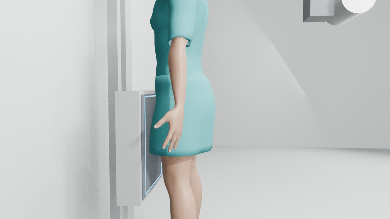
The routine abdomen views are Abdomen AP erect (acute abdomen), Abdomen AP erect (infant), Abdomen AP supine, Intravenous urography (IVU) series and Urinary tract survey (KUB). Abdomen lateral decubitus, Abdomen lateral erect, Abdomen PA erect, Pregnancy lateral erect and Pregnancy PA prone come in when you need to answer a particular question, or when the patient can't manage a routine view. Each guide walks you through the set-up step by step, with a short video, exposure settings for different patient sizes and what a good image should show.
A radiographer is still checking these guides.
Routine views
Extra views
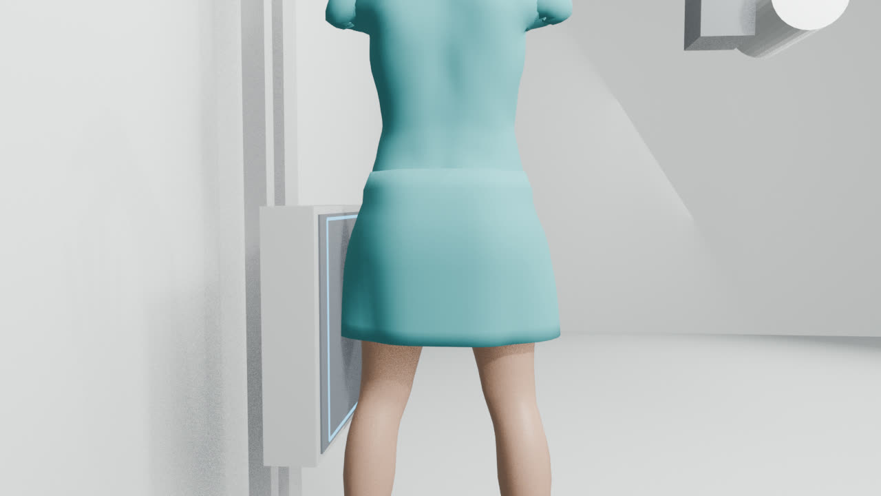
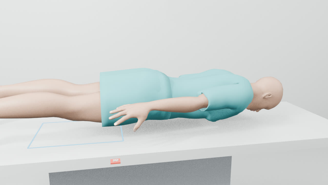
All views in this region
- Abdomen AP erect (acute abdomen)
- Abdomen AP erect (infant)
- Abdomen AP supine
- Intravenous urography (IVU) series
- Urinary tract survey (KUB)
- Abdomen lateral decubitus
- Abdomen lateral erect
- Abdomen PA erect
- Pregnancy lateral erect
- Pregnancy PA prone
Quick reference
Where to centre, how far and which receptor, for every abdomen view at a glance. Open a guide for the full set-up.
| Projection | Centre on | SID | Receptor | Breathing |
|---|---|---|---|---|
| Abdomen AP erect (acute abdomen) | the midline, high enough to include both domes of the diaphragm | 110 cm | 35×43 portrait, grid | Suspend respiration |
| Abdomen AP erect (infant) | the umbilicus | 110 cm | 24×30 portrait, grid | No breathing instruction needed |
| Abdomen AP supine | the midline at the level of the iliac crests | 100 cm | 35×43 portrait, grid | Breathe out and hold |
| Intravenous urography (IVU) series | the midline at the iliac crests | 100 cm | 35×43 portrait, grid | Breathe out and hold |
| Urinary tract survey (KUB) | the midline at the iliac crests | 100 cm | 35×43 portrait, grid | Breathe out and hold |
| Abdomen lateral decubitus | the midline, upper abdomen | 110 cm | 35×43 landscape, grid | Breathe out and hold |
| Abdomen lateral erect | the mid-coronal line at the iliac crest | 110 cm | 35×43 portrait, grid | Suspend respiration |
| Abdomen PA erect | the midline, including both domes of the diaphragm | 110 cm | 35×43 portrait, grid | Suspend respiration |
| Pregnancy lateral erect | the pelvis, including the symphysis | 110 cm | 35×43 portrait, grid | Suspend respiration |
| Pregnancy PA prone | the abdomen midline | 100 cm | 35×43 portrait, grid | Suspend respiration |
Mistakes that lead to repeats
- Free gas missed on the abdomen AP erect (acute abdomen): centre higher; the diaphragm matters more than the pelvis on this view.
- Diaphragm cut off on the abdomen AP erect (infant): centre at the umbilicus and open the field upward.
- Diaphragm not shown on the abdomen AP supine: recentre higher or add an upper film.
- Ureters not filled on the intravenous urography (IVU) series: take a prone film at 25 minutes if the doctor asks.
- Upper renal poles cut off on the urinary tract survey (KUB): recentre; take a second film if the abdomen is long.
- Up-side flank cut off on the abdomen lateral decubitus: shift centring toward the uppermost flank.
Common questions
- Which abdomen views are routine?
- Usually Abdomen AP erect (acute abdomen), Abdomen AP erect (infant), Abdomen AP supine, Intravenous urography (IVU) series and Urinary tract survey (KUB).
- What distance (SID) do abdomen X-rays use?
- Anywhere from 100 to 110 cm, depending on the view. Each guide gives the exact figure.
- Do abdomen X-rays need a grid?
- Yes, every view here is taken with a grid.
Try it in ScanFlo: the ScanFlo XR X-ray Room, coming soon, lets you set up every abdomen projection in a 3D room and score your positioning. See ScanFlo XR →