
Forearm X-ray positioning
How to position for both forearm X-ray views: where the patient goes, where to centre, which receptor and distance to use, and what a good image looks like.
By the ScanFlo Editorial Team · Updated
In this section · X-ray positioning
- X-ray positioning guides
- Skull positioning
- Face & sinuses positioning
- Soft tissue neck positioning
- C-spine positioning
- Chest positioning
- Shoulder positioning
- Humerus positioning
- Elbow positioning
- Forearm positioning
- Wrist positioning
- Hand positioning
- T-spine positioning
- Abdomen positioning
- L-spine positioning
- Sacrum & SI joints positioning
- Pelvis positioning
- Hip positioning
- Femur positioning
- Knee positioning
- Tibia & fibula positioning
- Ankle positioning
- Foot positioning
X-ray positioning/Forearm
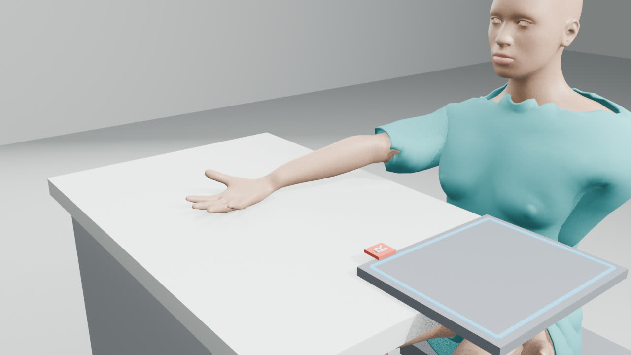
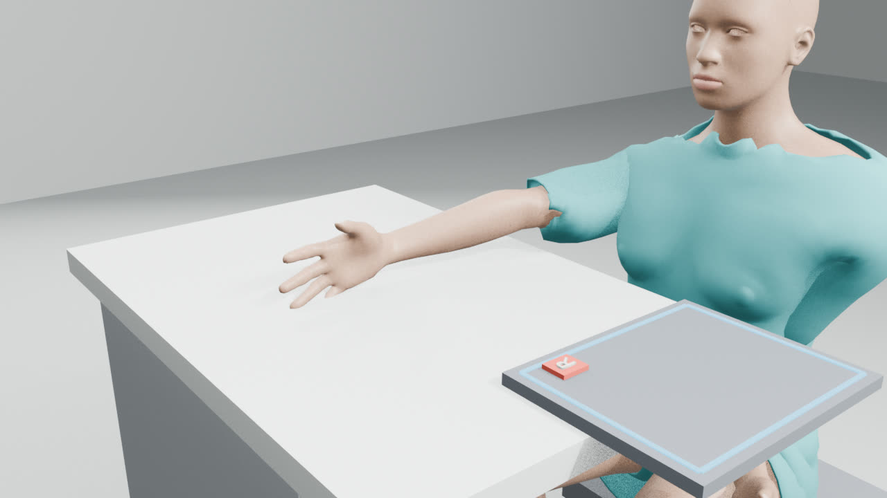
The routine forearm views are Forearm AP and Forearm lateral. Each guide walks you through the set-up step by step, with a short video, exposure settings for different patient sizes and what a good image should show.
A radiographer is still checking these guides.
Routine views
All views in this region
- Forearm AP
- Forearm lateral
Quick reference
Where to centre, how far and which receptor, for every forearm view at a glance. Open a guide for the full set-up.
| Projection | Centre on | SID | Receptor | Breathing |
|---|---|---|---|---|
| Forearm AP | the mid-forearm | 100 cm | 24×30 portrait | No breathing instruction needed |
| Forearm lateral | the mid-forearm | 100 cm | 24×30 portrait | No breathing instruction needed |
Mistakes that lead to repeats
- Bones crossed on the forearm AP: supinate the hand.
- Not lateral at the wrist on the forearm lateral: thumb straight up.
Common questions
- Which forearm views are routine?
- Usually Forearm AP and Forearm lateral.
- What distance (SID) do forearm X-rays use?
- 100 cm for every forearm view here.
- Do forearm X-rays need a grid?
- No. These parts are thin enough that a grid isn't needed.
Try it in ScanFlo: the ScanFlo XR X-ray Room, coming soon, lets you set up every forearm projection in a 3D room and score your positioning. See ScanFlo XR →