
Reconstruction, kernels and windows
One CT acquisition can become a soft-tissue series, a lung series and a bone series. The kernel, slice thickness and window decide which, and all three are chosen after the photons are already collected.
By the ScanFlo Editorial Team · Updated
In this section · CT fundamentals
A CT acquisition does not produce an image. It produces projections: attenuation measured from hundreds of angles around the patient. The image you read is one of many that could be built from those measurements. Protocol basics lists the parameters; the aim here is to explain why the same raw data can give such different pictures.
Hounsfield units: the number under every pixel
Every reconstructed voxel holds an attenuation value on a fixed scale. Water is defined as 0 HU and air as −1000. Fat sits around −100, soft tissue a few tens above zero, and bone runs into the hundreds and beyond. Because the scale is fixed, the numbers mean something: a lesion measuring −90 HU contains fat, and a fluid collection that measures 40 HU is not simple fluid.
The kernel: sharpness against noise
Reconstruction applies a filter, called the kernel, to the projections before back-projecting them into the image. A soft kernel lets through mostly low spatial frequencies. Edges are gentle and noise is low, which is what soft tissue needs: a small difference in density against a quiet background. A sharp kernel lets high frequencies through. Fine structures such as bronchial walls, trabeculae and the ossicles become crisp, but the noise is amplified along with them.
Neither is better. They answer different questions from the same photons. That is why a chest CT usually leaves the console as a soft-tissue series and a lung series, and a trauma spine as soft tissue and bone. On the consoles ScanFlo follows, kernel names read as a letter and a number: B for body and H for head, with higher numbers sharper. A B30 is a soft body kernel; a B70 is a sharp one.
Iterative reconstruction, now standard on most scanners, models noise and the scanner geometry and refines the image over several passes. It lowers noise at a given dose, which is what makes lower-dose protocols usable. At high strength it can give soft tissue a smooth, slightly plastic texture that some readers dislike.
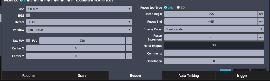
Slice thickness and increment
Thin slices keep z-axis detail, show small nodules without partial-volume blurring and feed good reformats. They are also noisier, because each slice holds fewer photons. Thick slices average that noise away and are faster to review, but a 3 mm nodule in a 5 mm slice is diluted by its neighbours.
The usual answer is to reconstruct thin for the archive and for reformats, and thick (or thin-overlapping, then reformatted) for primary reading. The increment, the spacing between reconstructed slices, can be smaller than the thickness. Overlapping slices improve reformats and lesion detection at no dose cost.
The window: a second choice, made at the viewer
The kernel is baked into the series. The window is not. A CT image holds far more grey levels than a monitor or an eye can separate, so the viewer maps a chosen band of HU onto black-to-white. The level sets the centre of that band; the width sets how wide it is. Everything below the band is black, everything above is white.
| Window | Width / Level (HU) | For |
|---|---|---|
| Brain | ~80 / 40 | Grey-white differentiation, early infarct |
| Soft tissue | ~350–400 / 40–50 | Abdomen, mediastinum |
| Lung | ~1500 / −600 | Parenchyma, airways |
| Bone | ~1800–2000 / 400–500 | Cortex, trabeculae, fractures |
Kernel and window work together. A lung window on a soft-kernel series is usable but blurred; a soft-tissue window on a sharp-kernel series looks grainy. Match the pair to the question.
Try it in ScanFlo: Scan the chest case and reconstruct the same acquisition twice from the Recon tab: a soft kernel such as B30 at 5 mm and a sharp kernel such as B60 at 1 mm. View each with the lung and mediastinal presets. Then measure a region of the aorta on both series and compare the noise. See ScanFlo CT →