
ScanFlo PET-CT
The PET-CT simulator, from topogram to SUV.
Plan the PET and the CT on one topogram, lay out the beds, run the CT-AC and the PET, then read the fused study and measure it. These are the decisions a PET-CT technologist makes, on real DICOM cases, with the consequences on screen.

Definition
What is a PET-CT simulator?
A PET-CT simulator for technologist training reproduces the hybrid exam without a tracer or a scanner. You register the patient's weight, height, glucose and injected activity, plan the PET beds and the attenuation-correction CT on one topogram, run the acquisition, then read the fused images and measure SUV. ScanFlo PET-CT does this in a browser on real DICOM cases.
Coverage
What learners can drive
- Clinical PET-CT exam workspace with one exam chronicle: topogram, CT-AC, PET
- PET and CT ranges on one topogram with Match CT Range; step-and-shoot beds with a time per bed, or continuous bed motion
- CT reconstruction jobs with an explicit AC-CT feeding the PET; PET jobs with iterations, subsets, filter and matrix
- FDG registration (weight, height, glucose, activity, injection time), paediatric weight-based dosing, uptake clock, SUVbw and SUVlbm
- Fused axial, coronal and sagittal MPR with blend, PET palettes and SUV windows; rotating PT MIP, CT MIP and VRT; registration QC with a manual rigid nudge
- VOI SUVmax, SUVmean, SUVpeak, MTV and TLG; liver and blood-pool references, PERCIST threshold and Deauville; Lesion Scout, RECIST and HU ROI; timepoint comparison
- MI Neurology (normal-database z-scores) and MI Cardiology (17-segment polar maps) with dynamic and ECG-gated cardiac NH3 protocols
- Real DICOM PET-CT studies: FDG, PSMA, NaF, DOTATATE, cardiac NH3 and brain FDG, with a patient browser and filming
Inside the console
Real screens, not renders.
Every image here is a capture from the simulator learners actually use.
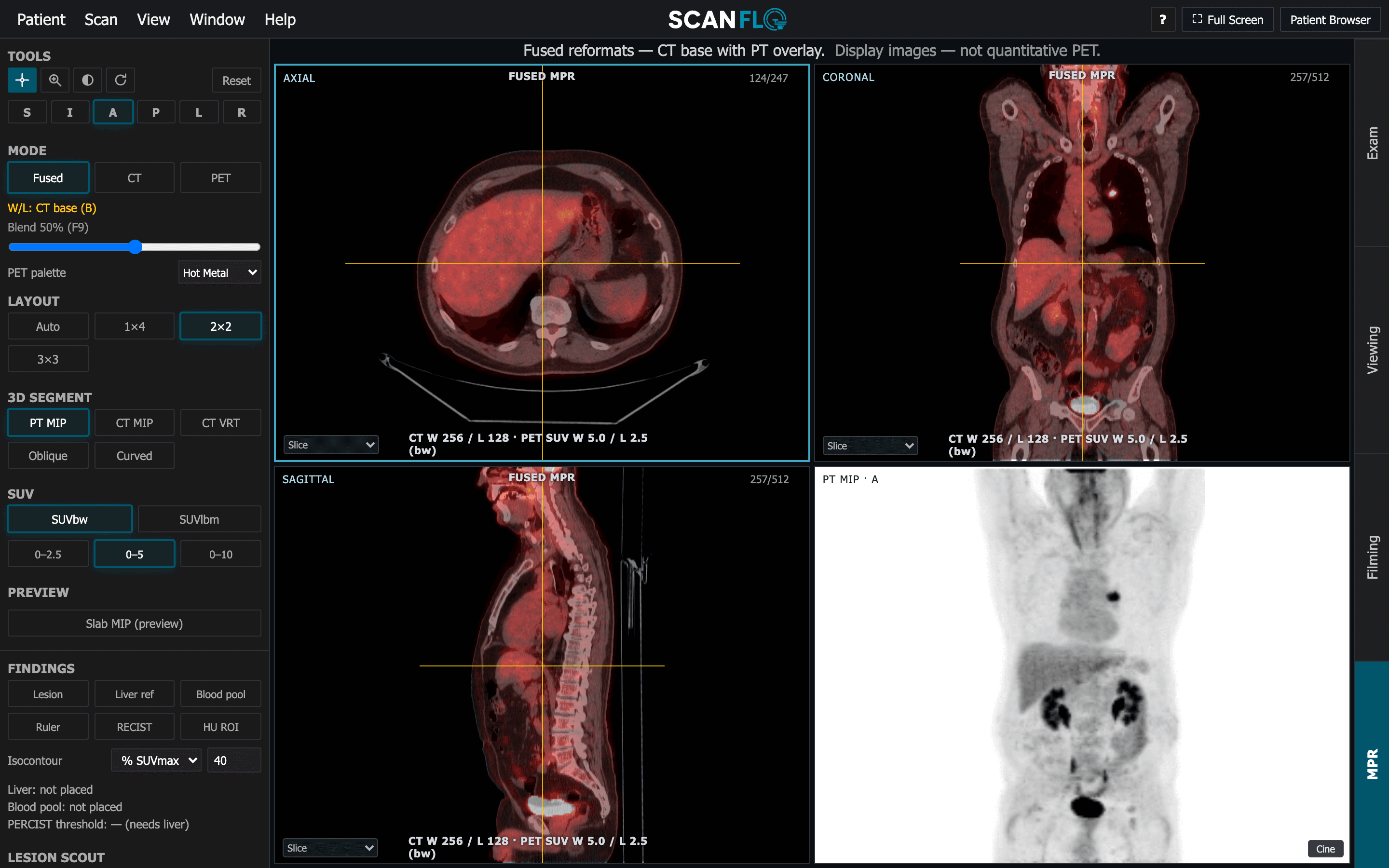
ScanFlo PET-CT: MPR · Fused reformats + PT MIPMPR: fused axial, coronal and sagittal reformats with blend and palette, the rotating PT MIP, SUV windows and findings tools. ScanFlo PET-CT: Exam · Whole-body FDG, 5 bedsExam workspace: topogram, CT-AC and PET in one chronicle, with beds planned on the topogram and a time per bed.
Workflow realism
One chronicle, one topogram, every bed.
The exam runs the way a clinical PET-CT console runs it: the topogram first, then the CT-AC, then the PET, all in one chronicle. The PET range is planned on the topogram and the CT follows it with Match CT Range, so the attenuation CT covers the PET field. The beds are laid out on the same image, each with its own time.
- 01Topogram → CT-AC → PET in a single exam chronicle
- 02PET and CT ranges planned together; Match CT Range makes the CT follow the PET
- 03Step-and-shoot with a duration per bed, overlap and total time
- 04Continuous bed motion as the alternative to beds
Physics by doing
Every SUV has a registration behind it.
SUV is only as good as what was entered at the desk. Learners register the patient (weight, height, glucose, activity and injection time), watch the uptake clock run and then read SUVbw and SUVlbm from those entries. The CT-AC's reconstruction jobs include an explicit AC-CT, and the PET reconstruction's iterations, subsets, filter and matrix are theirs to set.
- 01FDG registration with paediatric weight-based dosing
- 02Uptake clock from the injection time
- 03SUVbw and SUVlbm read from the registered values
- 04An explicit AC-CT feeding the PET reconstruction; PET jobs with iterations, subsets, filter and matrix
Reading and measuring
Read it fused, then put numbers on it.
The fused study opens in axial, coronal and sagittal planes with a blend, PET palettes and SUV windows beside a rotating PT MIP. Registration QC checks the PET against the CT, with a manual rigid nudge when they disagree. Then the findings: VOIs with SUVmax, SUVmean, SUVpeak, MTV and TLG, liver and blood-pool references for PERCIST and Deauville, and a second timepoint to compare against.
- 01Fused MPR with blend, palettes and SUV windows; PT MIP, CT MIP and VRT
- 02Registration QC with a manual rigid nudge
- 03VOI quantification, Lesion Scout, RECIST and HU ROI measurements
- 04Liver and blood-pool references, PERCIST threshold, Deauville, timepoint comparison
Try it on the console yourself.
Go deeper
Every capability, explained properly.
How each part of the console works, what it teaches, and how programs use it.
FAQ
Frequently asked questions
Is ScanFlo PET-CT available now?
Yes. ScanFlo PET-CT runs in the browser today, with Student and Student Pro plans on the pricing page.
Are the images real or generated?
The cases are real DICOM PET-CT studies (FDG, PSMA, NaF, DOTATATE, cardiac NH3 and brain FDG), opened from a patient browser the way a department workstation opens them.
Which scanner workflow does the console follow?
The exam workspace follows clinical PET-CT consoles: one exam chronicle from topogram to CT-AC to PET, range planning on the topogram, step-and-shoot beds or continuous bed motion, and separate CT and PET reconstruction jobs.
What quantification does it teach?
SUVbw and SUVlbm; VOI SUVmax, SUVmean, SUVpeak, MTV and TLG; liver and blood-pool references with the PERCIST threshold and Deauville scoring; RECIST and HU ROI measurements on the CT; and comparison between timepoints.
Does it cover neurology and cardiology as well as oncology?
Yes. MI Neurology compares brain FDG against a normal database as z-scores, and MI Cardiology shows perfusion on 17-segment polar maps from dynamic and ECG-gated cardiac NH3 protocols.
Start today
Put your learners at the console today.
Free 45-minute trial. No installs. Runs in any browser.