
CT scan simulators for students
Training software that reproduces a diagnostic CT console: topogram, kV, mAs, pitch, contrast timing, reconstruction and dose. How it differs from a radiotherapy CT simulator, and what the evidence says.
By the ScanFlo Editorial Team · Updated
In this section · Simulation
A CT scan simulator for students is software that reproduces a diagnostic CT console. You plan the scan range on a topogram, set kV, mAs, pitch and slice thickness, time the contrast, reconstruct with different kernels, and see what each choice does to image noise and to dose (CTDIvol and DLP), without a scanner or radiation. It is not the radiotherapy "CT simulator", which is a real scanner used to plan treatment.
Sources: Paolicchi et al. (2016); Gunn et al. (2021); Malik et al. (2024).
CT scan simulator or CT simulator: which do you mean?
Two different things share the name. Diagnostic radiography students want training software; radiotherapy departments mean a scanner.
Two meanings of 'CT simulator'
Radiotherapy centres describe CT simulation as "a specialized imaging procedure used to precisely plan radiation therapy treatments" (Revere Health), and the AAPM's Task Group 66 report sets out quality assurance for those scanners (Mutic et al., 2003). Everything below is about the training kind.
What does a CT scan simulator teach?
The decisions a CT technologist makes on every scan, in the order they make them.
One CT exam on a simulator
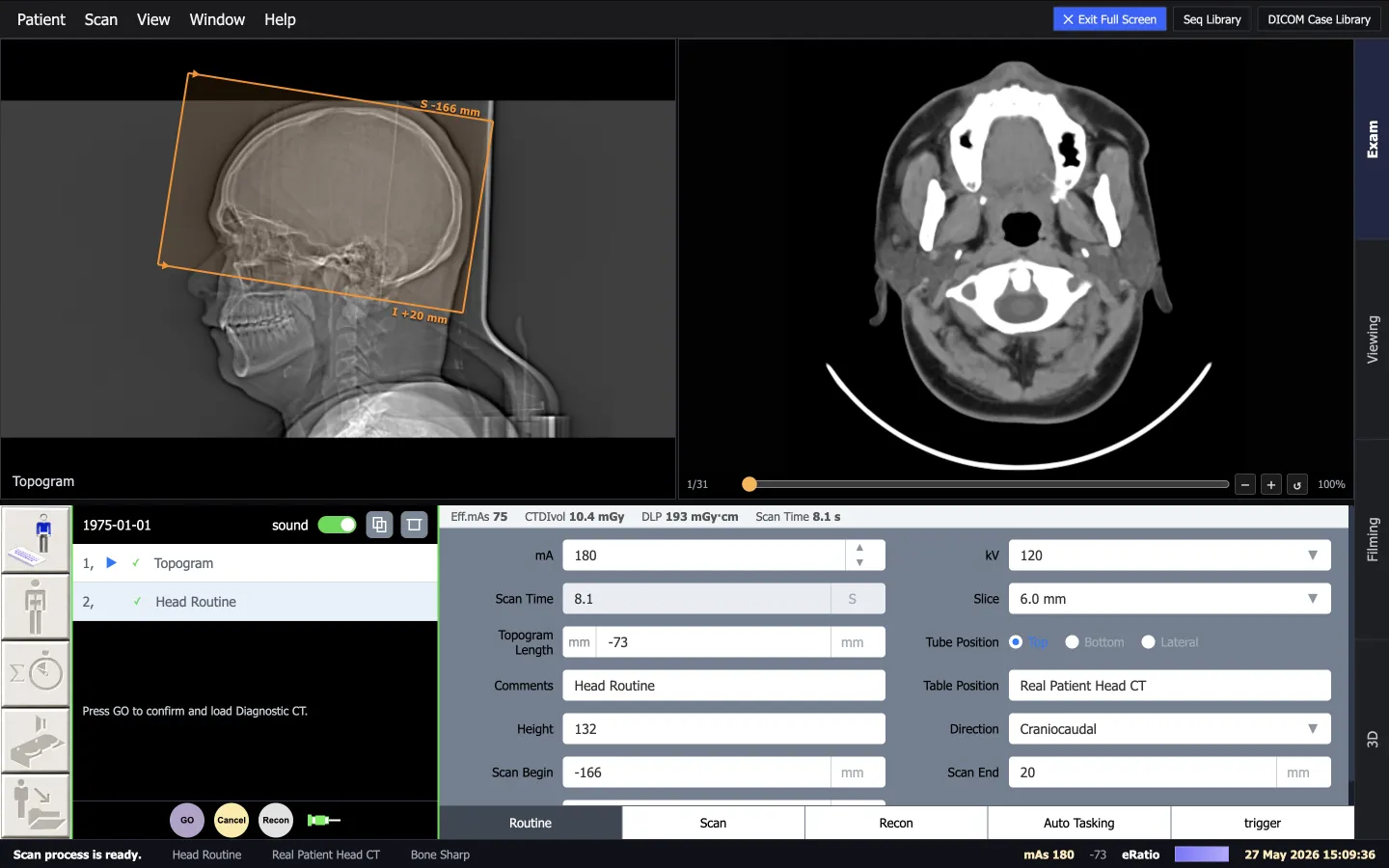
- STEP 01TopogramAcquire the localizer and set the start and end of the range.
- STEP 02kV and mAsTube potential and tube current-time product, or automatic exposure control.
- STEP 03Pitch and sliceTable travel per rotation against beam width; collimation and thickness.
- STEP 04Contrast timingBolus tracking: a region of interest and an HU threshold that triggers the scan.
- STEP 05ReconstructionKernel and window: soft tissue, lung or bone from the same raw data.
- STEP 06Dose readoutCTDIvol and DLP after every scan, against a reference level.
| Term | What it means |
|---|---|
| Topogram | The localizer radiograph used to prescribe the start and end of the scan range (also called scout, scanogram or surview). |
| kV | Tube potential: the voltage that accelerates electrons towards the target. |
| mAs | Tube current-time product per rotation. |
| Pitch | Table travel per rotation divided by total nominal beam width. |
| AEC | Automatic exposure control: adapts tube current to patient size for a set image quality. |
| Kernel | The reconstruction property that sets sharpness or smoothness in the axial plane. |
| Bolus tracking | Starts the scan automatically when contrast in a region of interest reaches a threshold. |
Pitch is a good example of what a simulator makes visible. Radiopaedia summarises the trade-off: a pitch above 1 lowers dose but also image quality, while a pitch below 1 improves image quality at a higher dose (Radiopaedia, pitch). On a simulator you change it and watch both happen.
ScanFlo CT · exam cockpit
Why dose teaching matters
Because many people who order and run CT scans underestimate the dose, and the console numbers are easy to misread.
Radiation protection: confidence vs practice
Read the console numbers carefully. CTDIvol is CTDIw divided by pitch, and DLP is CTDIvol multiplied by scan length, but neither is the patient's actual absorbed or effective dose (Radiopaedia, CT dose index). In India, AERB's safety code requires the CT console to display and record CTDI, DLP and exposure parameters, and asks practitioners to stay in line with international reference levels or those the regulator recommends (AERB RF-MED/SC-3).
Head CT, CTDIvol (mGy): Indian surveys vs an international figure
Does CT simulation training work?
Early studies are encouraging on knowledge and confidence, and they show transfer depends on how close the simulator is to the scanner students meet.
Students' learning can be optimised by repetitive practice in a safe environment and can be associated with increase student clinical confidence.
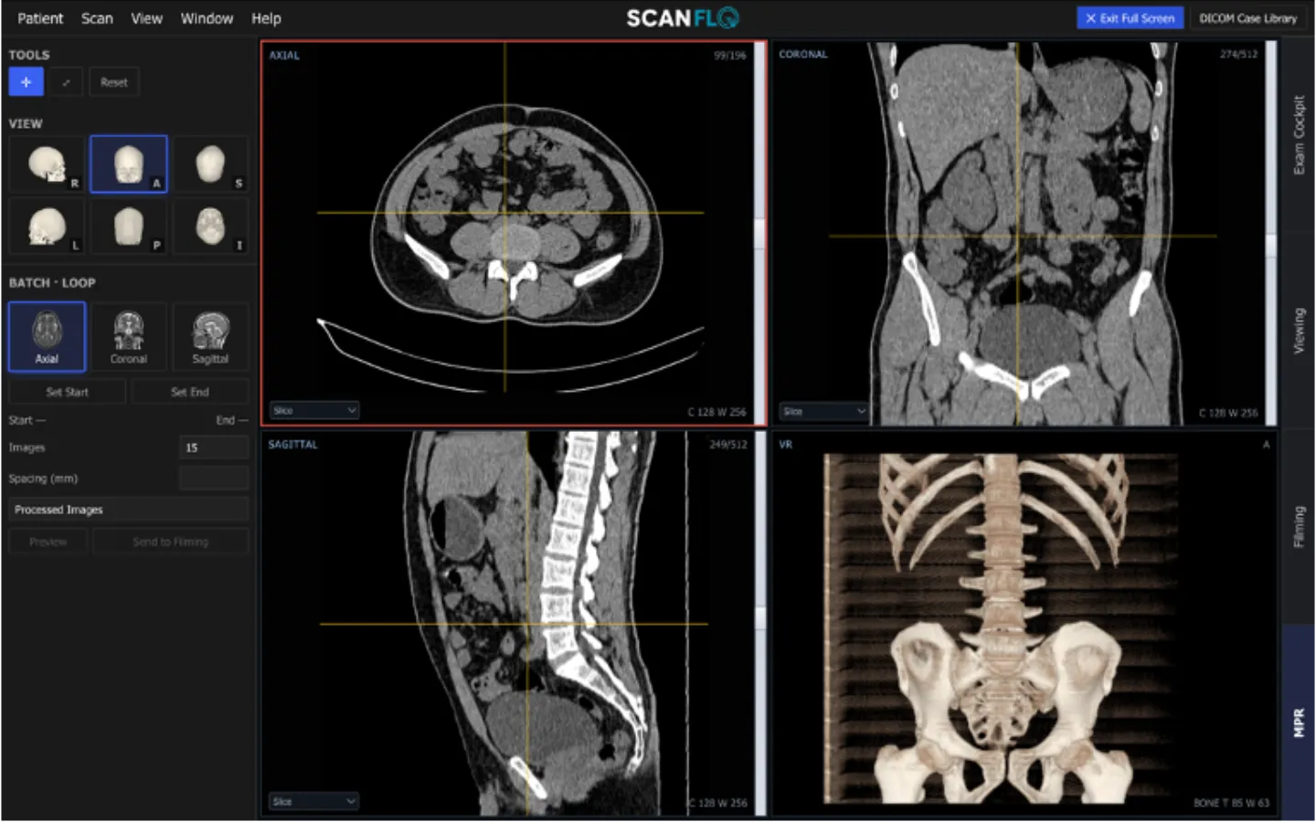
ScanFlo CT · MPR and 3D
What to look for in CT training software
Try it on the console
Try a CT scan simulator in your browser
ScanFlo CT: plan on the topogram, set the protocol, time the contrast and watch CTDIvol and DLP respond.
Start free →Related: CT dose metrics, contrast timing and bolus tracking, and the radiography simulation guide.
Frequently asked questions
Is a CT scan simulator the same as a CT simulator in radiotherapy?
No. In radiotherapy, a CT simulator is a real CT scanner used to image patients in treatment position so physicists can plan radiation beams. A CT scan simulator for students is training software that reproduces a diagnostic CT console without a scanner or radiation.
Does CTDIvol tell me the patient's dose?
Not exactly. CTDIvol and DLP describe the scanner's output measured in a standard phantom. They are the right numbers to compare protocols and check against reference levels, but they are not the absorbed or effective dose for an individual patient.
Does India have national CT diagnostic reference levels?
A 2018 study in the Indian Journal of Radiology and Imaging stated that India did not yet have national or region-specific CT reference levels. Regional surveys have proposed values since, and AERB's safety code asks practitioners to stay in line with international reference levels or those the regulator recommends.
Can I learn contrast timing on a simulator?
Yes, the logic of it: when to start monitoring, where to place the region of interest, which threshold in HU triggers the scan, and what happens to arterial and venous enhancement if you start too early or too late. Injecting a real patient stays a supervised clinical skill.
Do I need a VR headset for CT simulation?
No. A narrative review found immersive VR improved confidence and motivation, but noted it is still debated whether VR teaches CT better than computer-based simulation. A browser simulator on a lab computer covers the console decisions.
Sources
Every figure on this page links back to one of these.
- Mutic S, et al. Quality assurance for computed-tomography simulators and the computed-tomography-simulation process: AAPM TG-66. Medical Physics, 2003
- Revere Health. CT simulation
- AAPM. CT terminology lexicon, version 2.0, 2022
- Radiopaedia. Pitch
- Radiopaedia. CT dose index
- AERB. Safety code AERB/RF-MED/SC-3 (Rev. 2)
- Varghese B, et al. Radiation dose metrics in multidetector CT examinations: a multicentre study from seven tertiary care hospitals in Kerala. IJRI, 2018
- Saravanakumar A, et al. Establishment of CT diagnostic reference levels in select procedures in South India. IJRR, 2016
- Malik MMUD, et al. An analysis of computed tomography diagnostic reference levels in India compared to other countries. Diagnostics, 2024
- Paolicchi F, et al. Assessment of radiation protection awareness and knowledge about radiological examination doses among Italian radiographers. Insights into Imaging, 2016
- Singh P, et al. A prospective study assessing clinicians attitude and knowledge on radiation exposure to patients during radiological investigations. J Nat Sci Biol Med, 2015
- Lee JH, et al. Perception of radiation dose and potential risks of computed tomography in emergency department medical personnel. Clin Exp Emerg Med, 2015
- Gunn T, et al. The use of virtual reality computed tomography simulation within a medical imaging and a radiation therapy undergraduate programme. JMRS, 2021
- Lee K, et al. Computed tomography learning via high-fidelity simulation for undergraduate radiography students. Radiography, 2020
- Lee K, McInerney J. Transferability of learning from computed tomography simulation into clinical practice: student perspectives. Interactive Learning Environments
- Taylor B, McLean G, Sim J. Immersive virtual reality for pre-registration computed tomography education of radiographers: a narrative review. JMRS, 2023