
ScanFlo RadPath
The same disease, on CT and on MRI.
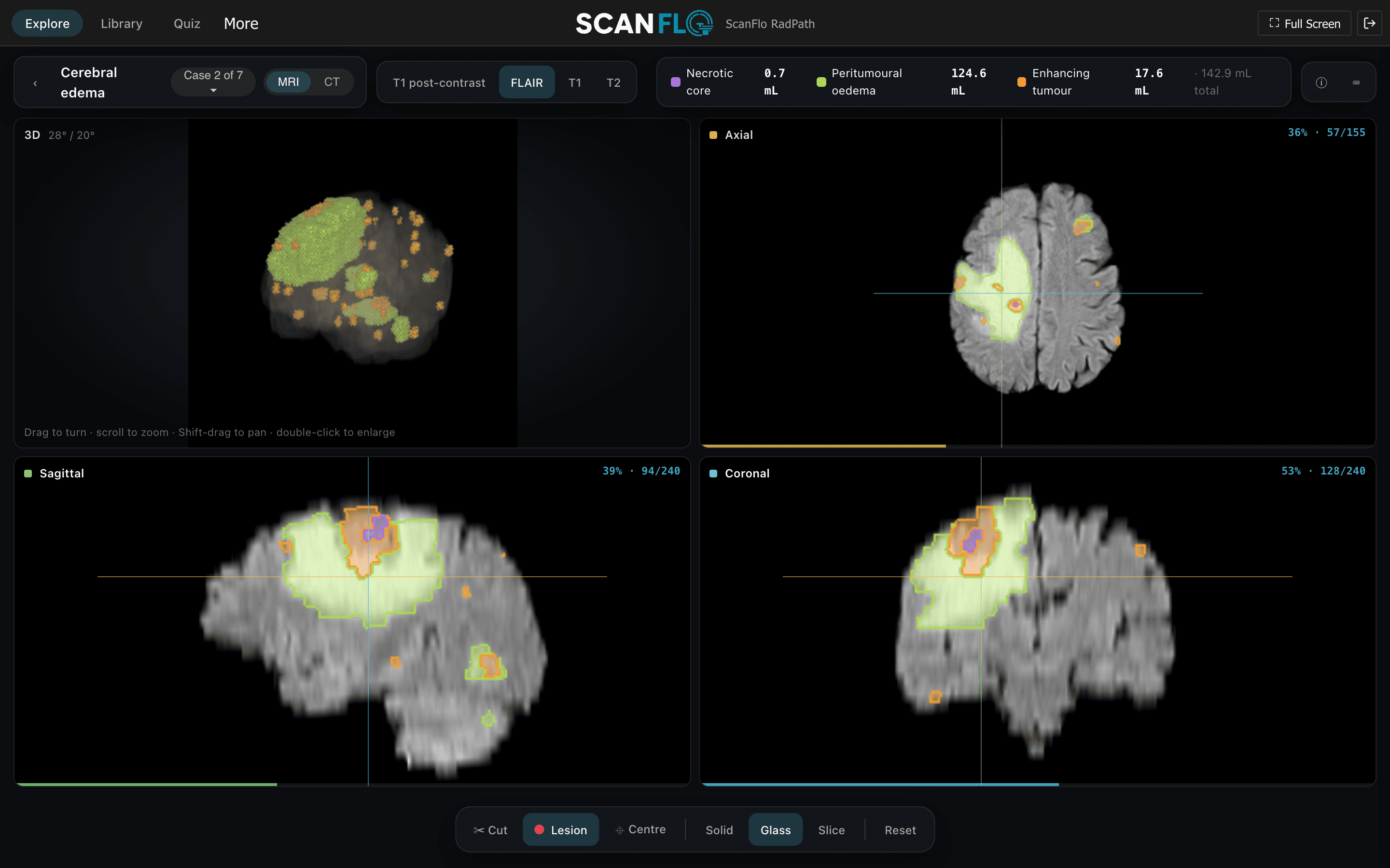
Pick a body region, open a condition such as a subarachnoid haemorrhage or a brain metastasis, and scroll through real cases with the lesion outlined, in 3D and in all three planes. Available now, alongside MRI, CT, PET-CT and Anatomy.
Coverage
Learn disease the way you will scan it.
- Brain, spine, thorax, abdomen, pelvis and the limbs, each with the conditions a radiographer meets most
- CT, MRI, PET-CT and X-ray studies from our own teaching collection and openly licensed datasets, each attributed
- Haemorrhage, infarct, oedema, tumour and fracture marked on the images, with the volume in mL
- Turn the case in 3D, cut into it, and scroll axial, sagittal and coronal side by side
- Switch modality and sequence on the same condition to see why each one shows it differently
- Runs in the browser on the same lab PCs that run the MRI and CT consoles
Open a case
The lesion, outlined and measured.
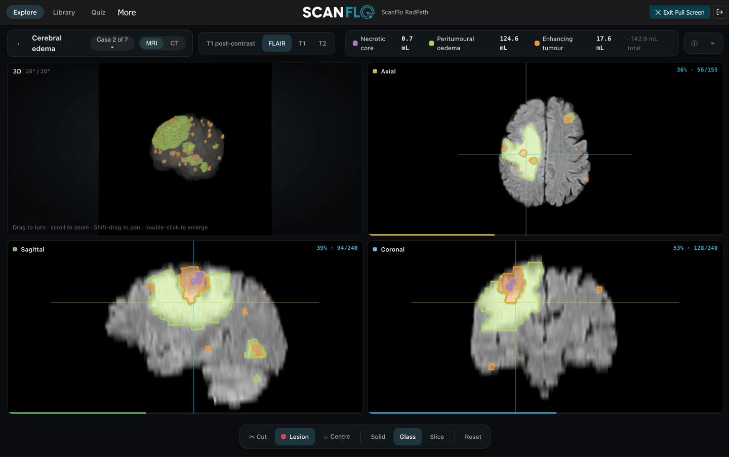
Open a case in 3D. The lesion is outlined in every plane, and each part of it is measured, so a learner sees where it is and how big it is before reading the images alone.
- 3D beside axial, sagittal and coronal
- Each component measured in mL
- The same condition on CT and on MRI
Find a case
Every case in one library.
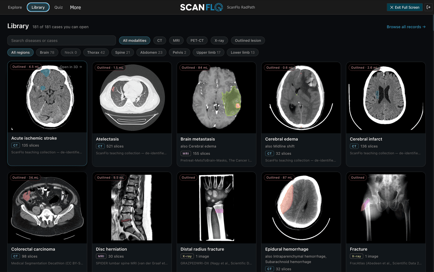
Filter by modality or region, and see the outlined volume before you open a case.
- Filter by modality and body region
- CT, MRI, PET-CT and X-ray
- Each case attributed to its source
Try it on the console yourself.
FAQ
Frequently asked questions
Are the RadPath cases real?
Yes. They are CT, MRI, PET-CT and X-ray studies from our own teaching collection and from openly licensed datasets, and each case is attributed.
What does RadPath show for each case?
The lesion outlined on the images (haemorrhage, infarct, oedema, tumour or fracture) in 3D and in axial, sagittal and coronal planes, with its volume in mL.
Does RadPath need a graphics card?
No. It runs in the browser on the same lab PCs that run the MRI and CT consoles.
Is RadPath a diagnostic tool?
No. RadPath is a teaching library for students and radiographers. It is not a medical device and is not for clinical or diagnostic use.
Start today
Start with RadPath today.
Trial it free, or pick a plan. Want a demo for your department? Talk to us.