
Fat suppression
Every fat suppression method has to tell a fat proton from a water proton. Fat sat uses frequency, STIR uses T1, Dixon uses phase, and each one fails exactly where its method breaks down.
By the ScanFlo Editorial Team · Updated
In this section · MRI fundamentals
Fat is bright on T1, bright on fast spin echo T2 and bright on most gradient echo sequences. That brightness hides oedema in bone marrow, flattens the conspicuity of enhancement and hides fluid in fat-rich regions such as the orbit. So fat suppression is routine, and choosing the method is a matter of knowing which one will hold up in the anatomy in front of you.
By frequency: fat sat
Fat protons precess slightly slower than water protons, about 3.5 parts per million, which is around 220 Hz at 1.5 T. Spectral fat saturation plays a narrow-band pulse tuned to the fat frequency, then a spoiler gradient, just before each excitation. Fat is left with nothing to give; water is untouched.
Frequency selection only works if the frequencies are where the scanner expects. Where the field is uneven, the fat peak shifts and the pulse misses it or hits water. The failures are familiar: bright fat at the edge of a large field of view, patchy suppression in the neck and around the shoulder, and suppression that collapses near metal. Good shimming and a centred region of interest help more than anything else.
By T1: STIR
Short tau inversion recovery ignores frequency. An inversion pulse flips everything, and excitation is timed for the moment fat, which recovers fastest, passes through zero. At 1.5 T that inversion time is roughly 150 ms, and longer at 3 T. Because it depends on T1 rather than frequency, STIR suppresses fat evenly across a large field of view and close to hardware. That is why it remains the default in marrow and after orthopaedic surgery.
It has two costs. Signal-to-noise ratio is lower, because every tissue is partly recovered at excitation. And STIR is not fat-specific: anything with a T1 like fat is nulled with it. Gadolinium shortens T1, so enhancing tissue can vanish on a post-contrast STIR. Do not use it after contrast.
Both at once: SPAIR
SPAIR uses an inversion pulse, like STIR, but one that is frequency-selective, like fat sat. Only fat is inverted, and excitation is timed for its null. The adiabatic pulse is less sensitive to RF inhomogeneity than a simple saturation pulse, so suppression is more uniform across the abdomen and breast. It still relies on the fat frequency, so metal and poor shimming still defeat it. SPIR is a closely related variant.
By phase: Dixon
Dixon does not suppress anything during acquisition. It reads echoes at times when fat and water are in phase and opposed (the same effect described in gradient echo), and then calculates a water-only image and a fat-only image. One acquisition gives four contrasts: in-phase, opposed-phase, water and fat. Because the calculation models field variation, Dixon holds up where fat sat fails, including large fields of view and many implants.
Its signature artefact is the fat-water swap: the calculation assigns a region to the wrong image, and the liver or a thigh suddenly looks like fat on the water image. It is obvious once you have seen it. Check the fat image whenever the water image looks strange.
| Method | Finds fat by | Strength | Fails when |
|---|---|---|---|
| Fat Sat | Frequency | Full SNR; fine after contrast | Field is uneven or near metal |
| STIR | T1 | Uniform, even near metal | Post-contrast; SNR matters |
| SPAIR | Frequency + T1 | Uniform over large FoV | Metal or poor shim |
| Dixon | Phase | Copes with uneven field; four contrasts | Fat-water swap |
Water excitation reaches the same goal from the opposite side, exciting only water protons. It is common in 3D cartilage imaging.
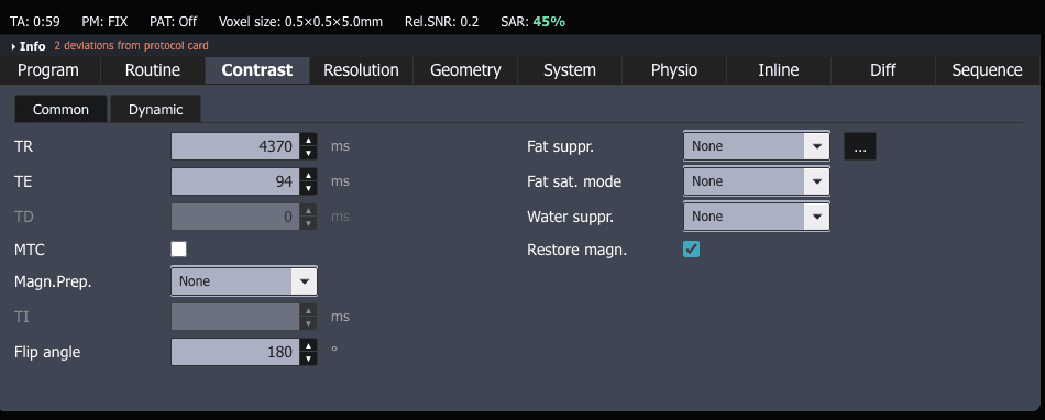
Try it in ScanFlo: Scan a knee with a PD or T2 sequence and set Fat suppr. to None, then Fat Sat. Look at the marrow, then compare with a STIR where the protocol offers one. On the post-contrast T1 of any protocol, note which options the console offers and why STIR is not among them. See ScanFlo MRI →