
Why practising on a real patient is the wrong way to learn protocols
ScanFlo Editorial Team · · 8 min read
In this article
You should not learn MRI protocols on real patients because every trial-and-error minute is paid for by someone lying in the bore: a repeated sequence, a longer exam, a heating risk, a delayed report. Learn the protocol logic first on a simulator, then refine your judgement on patients under supervision.
Nobody sets out to practise on a patient. It happens by default, when the first time a student plans a lumbar spine is the first time a real lumbar spine is on the table. The patient becomes the training material. There is a better order.
What does learning on the patient actually cost?
Time, money and comfort. One US study found that nearly one MRI exam in five needed at least one sequence repeated.
The same team put the lost revenue from motion at about $115,000 per scanner per year in their hospital, with a range of $92,600 to $139,000. A second, model-based study of brain MRI estimated $45,066 a year per scanner, rising to $364,242 when children were included (Slipsager and colleagues, 2020). Those are American-dollar estimates from rich-country hospitals, not Indian ones. The point carries anyway: repeats are expensive, and a nervous, slow operator makes them more likely.
Neuroradiologist Jalal Andre summed up his own data plainly in an interview: repeat sequences identified in nearly 20% of the MR examinations performed
(MedicalResearch.com, 2015). Every repeat is more time in the bore for someone who may already be in pain.
Can a protocol mistake injure a patient?
Yes. Most MRI injuries reported to the US FDA are burns, and many burns come down to how the patient was positioned and prepared.
The causes of those burns read like a positioning checklist. FDA reviewers traced them to a conductive object in the bore (16% of analysed reports), skin-to-skin contact forming a loop (16%) and skin touching the bore wall (10%). The loops they describe include crossed arms, touching thighs, touching calves and a thumb resting against the hip (Delfino, full text). The FDA adds that the potential for heating is greater during long MRI examinations
(FDA), which is one more reason a fumbling, slow protocol matters.
And the magnet itself is unforgiving. In January 2018 a man died at Nair Hospital in Mumbai after being pulled into an MRI scanner while holding an oxygen cylinder (The Quint, 2018). That was a screening failure, not a protocol error. It is the clearest reminder that MRI safety is learned before you need it.
What does WHO say about practising on patients?
WHO's patient safety curriculum says students should not perform a procedure on a patient for the very first time without preparation, and should first practise in a simulated environment.
It is very important that students not perform a procedure on a patient or administer a treatment for the very first time without appropriate preparation.
The guide lists screen-based computer simulators among the tools, and names their benefit simply: no risk to patients, and errors can be allowed to run their course. More than two decades ago Amitai Ziv and colleagues made the ethical argument in Academic Medicine: simulation tells everyone that patients are not commodities to be used as conveniences of training
(Ziv and colleagues, 2003).
Does learning on a simulator first actually work?
The broad evidence says yes. A meta-analysis of 609 studies found simulation training linked to large gains in knowledge and skills, and moderate gains for patients.
For MRI specifically the evidence is thinner. One randomised study of 60 students found those who used a simulator scored significantly higher than those who did not (Treceño-Fernández and colleagues, Sensors, 2021). It was a single day, and partly funded by a simulator maker, so treat it as encouraging rather than settled. The lead author of the big meta-analysis, David Cook of the Mayo Clinic, put the general picture this way: with extremely rare exceptions, we found improved outcomes for those who trained with simulation
(ScienceDaily, 2011).
What should you learn before the patient, and what only on the patient?
Protocol logic, planning and parameters can be learned beforehand. Communication, comfort and the unexpected need real people.
Learn on the patient
- Talking a frightened patient through the noise
- Real positioning, padding and coil placement
- Reading a patient's pain and deciding to cut a sequence
- Emergency response in the suite
Learn before the patient
- The order and purpose of each sequence in a protocol
- Planning slice groups on localizers
- What TR, TE, flip angle and matrix do to contrast and time
- Recognising and fixing common artefacts
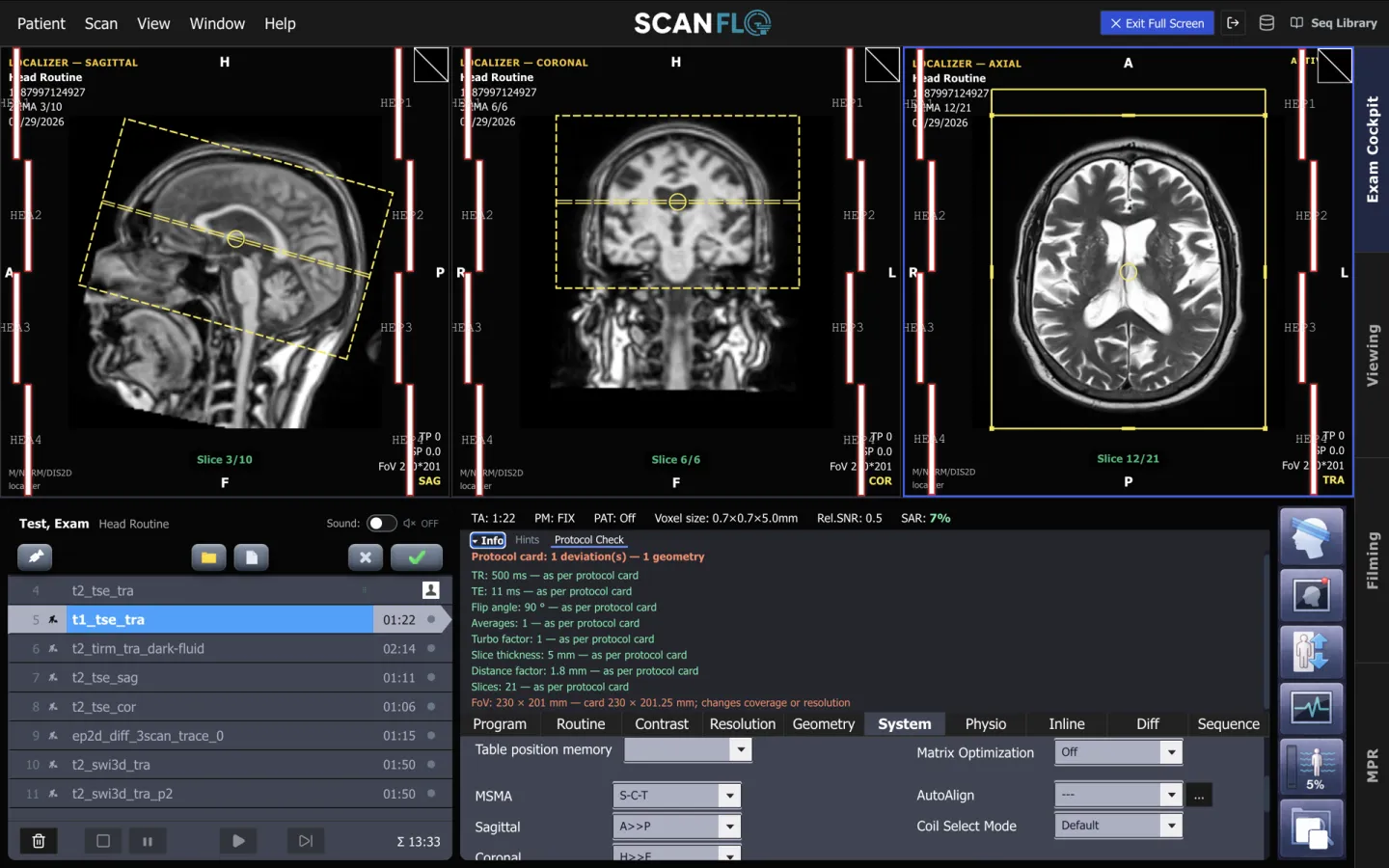
Try it on the console
Learn the protocol before the patient
Plan brain, spine and knee protocols on real localizers in ScanFlo MRI. Check your plan against the protocol card, then fix it.
Start free →Related: getting hands-on MRI practice before your first posting, whether simulator practice transfers to the scanner, and MRI protocol coverage.
Sources
Every figure in this article links back to one of these.
- World Health Organization. Patient safety fact sheet, 2023
- World Health Organization. Patient Safety Curriculum Guide: Multi-professional Edition, 2011
- Ziv A, Wolpe PR, Small SD, Glick S. Simulation-based medical education: an ethical imperative. Academic Medicine, 2003;78(8):783-8
- Andre JB, et al. Toward quantifying the prevalence, severity, and cost associated with patient motion during clinical MR examinations. JACR, 2015;12(7):689-95
- Slipsager JM, et al. Quantifying the financial savings of motion correction in brain MRI: a model-based estimate of the costs arising from patient head motion. JMRI, 2020;52(3):731-8
- Delfino JG, Krainak DM, Flesher SA, Miller DL. MRI-related FDA adverse event reports: a 10-yr review. Medical Physics, 2019;46(12):5562-71
- Delfino JG, et al. Full text of the 10-year review
- US Food and Drug Administration. MRI: benefits and risks
- Cook DA, et al. Technology-enhanced simulation for health professions education: a systematic review and meta-analysis. JAMA, 2011;306(9):978-88
- Mayo Clinic via ScienceDaily. Simulation an effective way to train health-care professionals, research review finds, 6 September 2011
- Treceño-Fernández D, et al. Magnetic resonance simulation in education: quantitative evaluation of an actual classroom experience. Sensors, 2021
- MedicalResearch.com. Patient motion during MRI scanning can lead to significant added costs (interview with Jalal Andre), 2015
- The Quint. Man gets sucked into MRI machine at Mumbai's Nair Hospital, dies, January 2018
Frequently asked questions
Isn't learning on real patients the only way to become a good technologist?
Real patients are essential, and nothing replaces supervised scanning. The point is the order. Protocol logic, planning and parameter choices can be learned beforehand, so supervised time on patients goes on judgement, communication and the surprises that only real people bring.
What causes most repeated MRI sequences?
Patient motion, in most published studies. Andre and colleagues found significant motion artefacts in 7.5% of outpatient and 29.4% of inpatient or emergency exams. Clear instructions, comfortable positioning and a shorter protocol all help, and a technologist who already knows the protocol runs it faster.
How do MRI burns happen?
Most often through radiofrequency heating: a conductive object in the bore, the patient's skin touching the bore wall, or skin-to-skin contact that forms a loop, such as crossed arms or touching thighs. FDA reviewers found these three made up a large share of burn reports.
What can I practise on a simulator?
Planning slice groups on localizers, choosing sequences for a clinical question, adjusting TR, TE, flip angle and resolution, recognising artefacts and fixing them, and timing a full protocol. Patient communication, screening conversations and real positioning still need people.