
Cancer in India: why early diagnosis matters, the growing role of PET-CT, and the need for skilled technicians
ScanFlo Editorial Team · · 13 min read
In this article
Two people can have the same cancer, in the same organ, with the same cells under the microscope, and face very different futures. Most of the time the difference isn't the hospital they reach or the drug they're given. It's when the disease was found.
In India it is usually found late. Registry data show that most breast, cervical and head and neck cancers are already locally advanced by the time they're diagnosed, and most lung cancers have already spread. That one fact shapes nearly everything that follows: which treatments are needed, what they cost a family, and how many people come through them.
This piece follows that thread through three parts of the system. How large the problem is. Why the timing of diagnosis matters so much, and where PET-CT fits in (and where it doesn't). And the part that gets least attention when a hospital announces a new scanner: the technologists who have to run it.
How many people, and which cancers
India's cancer numbers come from more than one source, and they don't agree to the last digit. The ICMR's National Cancer Registry Programme (NCRP) estimated 14,61,427 new cases for 2022. A 2025 analysis of 43 population-based registries by ICMR researchers, published in JAMA Network Open, projected 15,62,099 new cases and 8,74,404 deaths for 2024. WHO's Global Status Report on Cancer 2026 gives a similar case count for 2024 with a higher death estimate, 9,01,828.
The gaps come from method: which registries are used, how their rates are stretched over the many districts that have no registry, which year is modelled. For practical purposes the estimates tell one story. Around fifteen lakh Indians are diagnosed with cancer every year, and the number keeps going up. NCRP expects 2025 to be 12.8% higher than 2020.
On NCRP's numbers, about one Indian in nine will develop cancer before the age of 75. That average hides a lot. The 43-registry study put the national lifetime risk at 11.0% but found 21.1% in men and 18.9% in women in Mizoram, close to double.
Which cancers dominate depends on sex. In men the commonest are cancers of the mouth, lung and prostate. In women, breast cancer is far ahead of everything else (28.8% of female cases in NCRP's 2022 estimate), followed by the cervix and ovary. Several of these, the mouth, breast and cervix above all, give visible or palpable early signs and respond well to screening. Keep that in mind for the next section.
The real problem is timing
WHO's cancer fact sheet says it plainly: "When identified early, cancer is more likely to respond to treatment and can result in a greater probability of survival with less morbidity, as well as less expensive treatment." That's three separate benefits in one sentence. In India, all three are being lost at the same moment.
An NCRP analysis of hospital registry data (Mathur and colleagues, 2020) found that most patients with breast, cervical, head and neck and stomach cancer were diagnosed with locally advanced disease. For lung cancer, the commonest stage at diagnosis was distant metastasis.
Locally advanced doesn't mean hopeless. Many of these patients are still treated with the aim of cure. But the treatment is heavier: surgery, chemotherapy and radiotherapy together instead of one operation, longer courses, more side effects, more months away from work, and bills that many families pay out of their own pockets. Found a year earlier, the same cancer would often have been a smaller problem.
Why does it happen? The reasons are unglamorous. A review of cancer research in India in the Indian Journal of Medical Research lists them: low awareness among the public and among community doctors, few screening programmes, few diagnostic facilities close to home, long journeys to a tertiary cancer centre, cost, and the stigma that still attaches to the word cancer. None of these is a technology problem.
How much screening India does is genuinely disputed. The government's NP-NCD programme reports population-based screening for oral, breast and cervical cancers for people over 30, through primary health centres. WHO's 2026 report, which looks for organised, quality-assured programmes, lists India as having no programme for breast or colorectal screening and puts HPV-based cervical screening coverage at 0 to 9% of women aged 30 to 49. Both can be true. Screening is happening, but not yet at the coverage, and with the follow-up, needed to move the stage at which most cancers are found.
The national HPV vaccination programme launched in February 2026 is the most hopeful development here. Its effect on cervical cancer numbers will take years to show.
Where PET-CT fits, and where it doesn't
A common misunderstanding first. PET-CT is not a screening test. It isn't used to look for cancer in healthy people, and a clinic selling "full-body PET-CT check-ups" to people without symptoms is selling something the evidence doesn't back. The scan combines a radioactive tracer with a CT, it costs a lot, and in a symptom-free population the radiation, the expense and the false alarms outweigh what it finds.
PET-CT answers a different question: now that we know this person has cancer, where exactly is it, and is treatment working? The usual tracer is FDG, a glucose analogue labelled with fluorine-18. Many tumours burn glucose fast and show up as bright spots on the PET. The CT, taken in the same session, supplies the anatomy and corrects the PET for how much of the signal the body absorbed. Put together, a small node or a deposit in bone that looks innocent on CT alone can show up clearly.
That answer changes treatment. One of the largest studies of the effect, the US National Oncologic PET Registry (Hillner and colleagues, 2008), collected 22,975 scans and found that doctors changed their intended management after 36.5% of them. Those were older American patients, and a change of plan isn't the same thing as a better outcome. Still, the scale is hard to ignore. In more than a third of cases, the scan changed what was going to happen next.
| Cancer | Typical uses |
|---|---|
| Lymphoma | Initial staging of Hodgkin and aggressive non-Hodgkin lymphoma; interim and end-of-treatment response; suspected relapse |
| Lung (non-small cell) | Staging before surgery or radiotherapy, especially mediastinal nodes and distant spread; restaging; recurrence versus radiation change |
| Head and neck | Neck nodes with an unknown primary; staging; response after chemoradiation; recurrence when CT or MRI is equivocal |
| Breast (advanced) | Staging locally advanced or inflammatory cancer; looking for distant spread; suspected recurrence |
| Oesophagus and stomach | Staging before planned surgery; restaging after chemoradiation |
| Colorectal | Before surgery for metastases; rising CEA with negative conventional imaging |
| Cervix and ovary | Staging advanced cervical cancer; suspected recurrence, for example a rising CA‑125 |
| Melanoma | Staging high-risk disease for distant spread |
| Thyroid (differentiated) | Rising thyroglobulin with a negative radioiodine scan |
It has blind spots too. The normal brain uses a great deal of glucose, so FDG shows brain tumours poorly against that background, and MRI does that job. Most prostate cancers take up little FDG; PSMA tracers are used instead. Some slow-growing and mucinous tumours barely show. A good PET-CT report is read alongside the other imaging, not instead of it.
So PET-CT doesn't fix late diagnosis. What it does is help at the point where most Indian patients actually are. It stages a locally advanced cancer accurately, so a curative plan isn't wasted on someone whose disease has already spread, or withheld from someone whose hasn't. And it shows early whether treatment is working, while there's still time to change course.
The machines are arriving
Singh and colleagues, writing in the Indian Journal of Nuclear Medicine in 2023, counted 442 working nuclear medicine centres in India, with 359 PET-CT scanners and 24 medical cyclotrons. That is a long way from where the country was a decade ago.
For 1.4 billion people it is still thin. By our arithmetic, 359 scanners works out to roughly one for every four million Indians, about 0.25 per million. The USA and Japan have around 4 per million. Singh's group estimates India would need about 1,000 more scanners over the next ten years just to reach one per million, and WHO's 2026 report puts India in its lowest band, 0 to 1 PET scanner per million people.
Where the scanners are matters as much as how many there are. FDG has a half-life of about 110 minutes, so half of it is gone in under two hours. A scanner depends on a cyclotron close enough to get the tracer to it while it's still useful. With two dozen cyclotrons in the country, PET-CT naturally gathers around the cities that have one, and a patient from a high-burden district may be a long journey from the nearest scan.
The part nobody budgets for: people
A scanner purchase has a line in the budget. The people who run it often don't.
The same 2023 paper estimates that India trains about 150 nuclear medicine technologists a year (BSc and MSc graduates, from about 25 institutes), and around 50 nuclear medicine physicians. Set that against 442 centres and 359 scanners, each needing trained staff on every shift it runs, with more centres opening every year. Each new PET-CT recruits from the same small pool. We couldn't find a published national count of working nuclear medicine technologists, which is itself part of the problem: nobody is measuring the gap.
What does a PET-CT technologist actually do? Much more than press start.
None of these errors are exotic. A patient who had breakfast shows a bright heart and muscles, and less uptake in the tumour. A patient who chatted or chewed through uptake shows lit-up vocal cords and jaw muscles. A CT taken in a deep breath-in, against a PET acquired while breathing freely, can put a liver lesion's hot spot in the base of the lung on the fused image. These are the things a newly qualified technologist learns, often by getting one of them wrong on a real patient.
The training route is also changing. Nuclear medicine technology courses used to be assessed by AERB for recognition. That assessment has been discontinued, and institutes now approach the National Commission for Allied and Healthcare Professions (NCAHP). For students, that means checking a course's current status before enrolling rather than relying on an old list.
Training before the scanner
Nothing replaces supervised time in a working department with a real hot lab. But that time is scarce. The scanner is booked from morning to evening, the patient on the table has already been injected with radioactivity, and no department lets a student learn by trial and error on that list.
Simulation can take over the parts that don't need a patient: planning bed positions on a topogram, entering the dose and timing correctly, choosing a reconstruction, reading fused images, recognising misregistration when you see it. Those are the steps where a mistake costs nothing on a simulator and a repeat scan, or a wrong SUV, on a patient.
That's why we built a PET-CT simulator into ScanFlo, alongside our CT simulator and MRI simulator. The two screens below are from it.
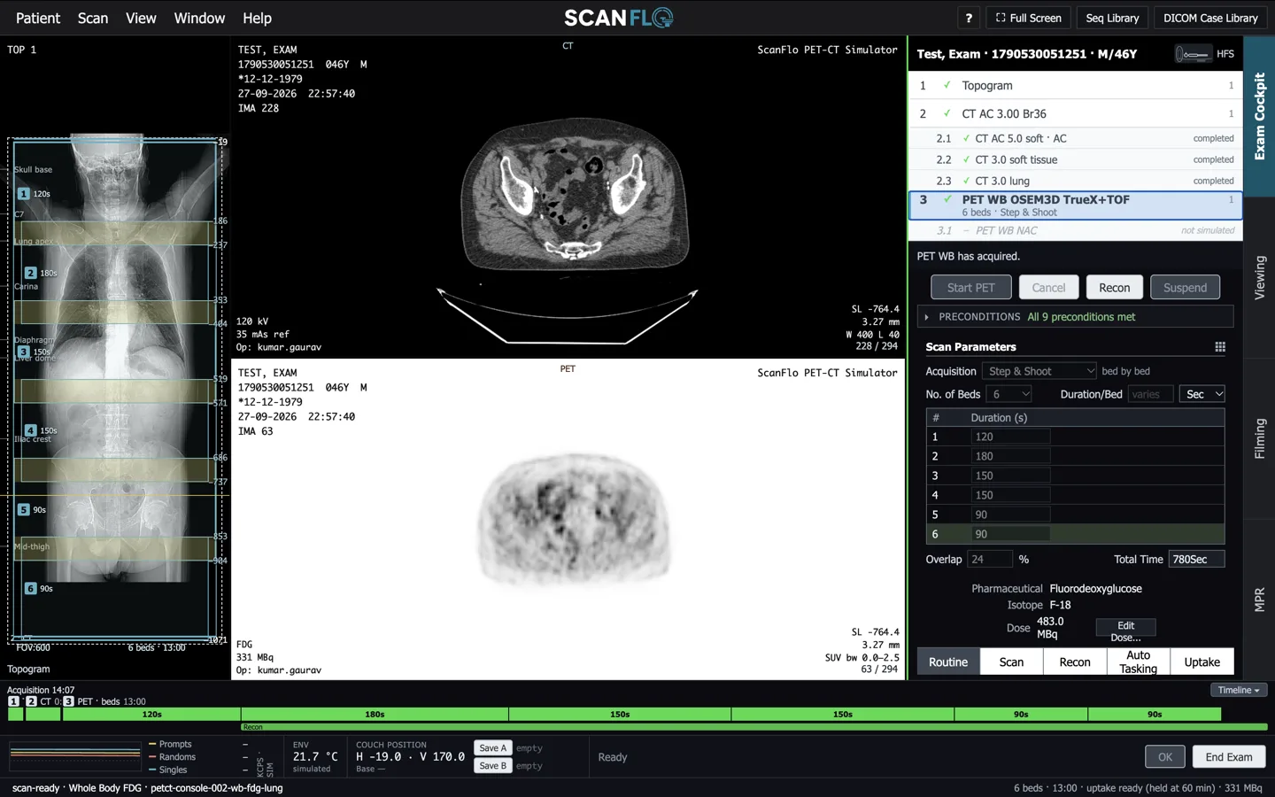
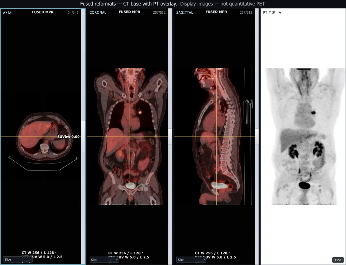
We should be clear about what it isn't. The images are display images for training, not quantitative PET, and a simulator doesn't teach dose dispensing or radiation protection in a hot lab. Those need the real thing, with a supervisor. What it does is let a student arrive at that first posting already knowing the order of the exam and what a bad fusion looks like, so the scarce hours in the department go on the parts only a department can teach. (The CT half of the exam carries its own dose questions; our piece on low-dose CT goes into those.)
What to take away
If you are a student
Nuclear medicine technology is a small field with a growing number of scanners to staff. Choose a course with current recognition and real PET-CT postings. Take patient preparation as seriously as the scanner, because that is where most spoiled scans start.
If you run a department
Budget the team when you budget the scanner. Plan who covers every shift, how new staff are trained before their first patient, and how you will check dose records and fusion quality. A scanner that runs short-staffed runs slower, and makes mistakes.
If you shape policy
Track stage at diagnosis as closely as cases. Put cyclotrons where the burden is, not only where the market is. And count the workforce: without a national figure for practising technologists, nobody can say how short we are.
India will keep adding PET-CT scanners, and it should. Whether they change outcomes depends on two things that don't arrive in the crate: patients who reach the scanner early enough for the result to matter, and technologists skilled enough to make every scan worth reading.
A note on figures. Case and death estimates come from different sources and years and are labelled with both; they are not directly comparable. Stage-at-diagnosis shares are from hospital-based registries, which see patients who reached a cancer hospital. Scanner, cyclotron and graduate numbers are from the 2023 review cited and will have grown since. The per-million India figure is our own arithmetic. The PET-CT uses in the table are common indications, not a clinical guideline. The pathway figure is an illustration. This article is general information, not medical advice.
Sources
- Sathishkumar K, Chaturvedi M, Das P, et al. Cancer incidence estimates for 2022 & projection for 2025: result from National Cancer Registry Programme, India. Indian J Med Res 2022;156(4&5):598-607. IJMR
- ICMR-NCDIR. Cancer incidence and mortality across 43 cancer registries in India. JAMA Netw Open 2025;8(8):e2527805. doi
- Mathur P, Sathishkumar K, Chaturvedi M, et al. Cancer statistics, 2020: report from National Cancer Registry Programme, India. JCO Glob Oncol 2020;6:1063-75. doi
- World Health Organization. Global status report on cancer 2026, and Kurian OC, Cancer care in India: reading the WHO Global Status Report 2026, Observer Research Foundation, 19 August 2026
- World Health Organization. Cancer fact sheet
- Cancer research in India: challenges and opportunities. Indian J Med Res. doi
- Singh B, Sahani G, Prasad V, Watts A, Kumar R. India's growing nuclear medicine infrastructure and emergence of radiotheranostics in cancer care. Indian J Nucl Med 2023. PMC
- Hillner BE, Siegel BA, Liu D, et al. Impact of PET/CT and PET alone on expected management of patients with cancer: initial results from the National Oncologic PET Registry. J Clin Oncol 2008;26:2155-61. doi
- Atomic Energy Regulatory Board. Nuclear medicine facilities and notices on recognition of nuclear medicine technology courses
Frequently asked questions
Is PET-CT used for cancer screening in India?
No, and it should not be. PET-CT is used after cancer is diagnosed or strongly suspected: to stage it, to check whether treatment is working, to look for recurrence and to plan radiotherapy. In people without symptoms the radiation dose, cost and false alarms outweigh the benefit. Screening in India is done with simpler tests, such as oral visual examination, clinical breast examination and cervical screening, under the NP-NCD programme.
How many PET-CT scanners does India have?
A 2023 review in the Indian Journal of Nuclear Medicine (Singh and colleagues) counted 442 operating nuclear medicine centres with 359 PET-CT scanners and 24 medical cyclotrons. That is roughly 0.25 scanners per million people. The same authors estimate India would need about 1,000 more scanners over ten years to reach one per million. The WHO Global Status Report on Cancer 2026 places India in its 0 to 1 PET scanner per million band.
Why are so many cancers in India diagnosed late?
Low awareness of early symptoms among the public and some community doctors, limited organised screening, few diagnostic facilities close to home, long travel to a cancer centre, cost, and the stigma a cancer diagnosis still carries. Registry data show most breast, cervical and head and neck cancers are already locally advanced at diagnosis.
What does a PET-CT technologist do?
Prepares the patient (fasting, blood glucose, history), measures and records the radioactive dose in a dose calibrator, manages the uptake period, runs the CT and the PET bed positions, reconstructs and checks the fused images, and keeps staff, patients and the public safe from radiation. Each step has errors that can spoil a scan without being obvious at the console.
How do I become a PET-CT or nuclear medicine technologist in India?
Through a BSc or MSc in Nuclear Medicine Technology, or an equivalent diploma such as DMRIT, from a recognised institute. AERB used to assess these courses; that assessment has been discontinued and institutes now approach the National Commission for Allied and Healthcare Professions (NCAHP) for recognition. Check a course's current recognition status before you enrol, and look for one with regular clinical postings in a department that runs PET-CT.